Review
. 2022 May 7;10(5):1086.
doi: 10.3390/biomedicines10051086.

Cathelicidin LL-37 in Health and Diseases of the Oral Cavity

Affiliations
Review

Cathelicidin LL-37 in Health and Diseases of the Oral Cavity

Joanna Tokajuk et al. Biomedicines. .

Abstract

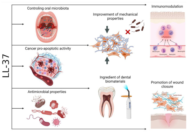
The mechanisms for maintaining oral cavity homeostasis are subject to the constant influence of many environmental factors, including various chemicals and microorganisms. Most of them act directly on the oral mucosa, which is the mechanical and immune barrier of the oral cavity, and such interaction might lead to the development of various oral pathologies and systemic diseases. Two important players in maintaining oral health or developing oral pathology are the oral microbiota and various immune molecules that are involved in controlling its quantitative and qualitative composition. The LL-37 peptide is an important molecule that upon release from human cathelicidin (hCAP-18) can directly perform antimicrobial action after insertion into surface structures of microorganisms and immunomodulatory function as an agonist of different cell membrane receptors. Oral LL-37 expression is an important factor in oral homeostasis that maintains the physiological microbiota but is also involved in the development of oral dysbiosis, infectious diseases (including viral, bacterial, and fungal infections), autoimmune diseases, and oral carcinomas. This peptide has also been proposed as a marker of inflammation severity and treatment outcome.

Keywords: antimicrobial peptides; human cathelicidin; immunomodulation; oral cavity.


Conflict of interest statement

The funders had no role in the design of the study; in the collection, analyses, or interpretation of data; in the writing of the manuscript; or in the decision to publish the results.

Figures

Figure 1
Figure 1
Expression of LL-37 in 35 human tissues, including oral epithelium, tonsils, and salivary glands (highlighted in orange). The data were obtained from ProteomicsDB (https://www.ProteomicsDB.org) last accessed 25 March 2022 [23] and represent mass spectrometry (MS1-level) proteome quantification; the intensity-based absolute quantification (iBAQ) value is a measure of protein abundance and corresponds to the sum of all the peptide intensities divided by the number of observable peptides of a protein (panel A). Relative abundance of CAMPs, including LL-37 (highlighted in red), present in the oral cavity (whole organism—integrated; LL-37 is highlighted in red). The data were obtained from PAXdb (Protein Abundance Database; https://pax-db.org/) last accessed 25 March 2022 ) [24] and are based on a mass-spectrometry-based study of the human proteome by Wilhelm et al. (2014) [25]; parts-per-million (ppm) values represent the abundance of each protein with reference to the entire expressed proteome, i.e., each protein entity is enumerated relative to all other protein molecules in the sample (panel B).
Figure 2
Figure 2
Tree map of the Biological Process (BP) Gene Ontology (GO) category for human cathelicidin LL-37 (panel A); green and red boxes show processes associated with immune responses against microorganisms and immunomodulatory action, respectively. Functional similarity of 20 human cationic antimicrobial peptides (CAMPs) present in the oral cavity inferred from comparing their Biological Process (BP) Gene Ontology (GO) category (panel B); the BPs unique for human cathelicidin LL-37 are indicated by yellow color in panel A. The dendrogram was constructed using Dice coefficient and UPGMA clustering with NTSYSpc software ver. 2.1 (Exeter Software) based on the GOs collected from the QuickGO server (www.ebi.ac.uk/QuickGO/).
Figure 3
Figure 3
Relative abundance of CAMPs, including LL-37 (highlighted in red), in whole organism (panel A) and saliva (panel B). The data were obtained from PAXdb (Protein Abundance Database; https://pax-db.org/, accessed on 25 March 2022) [24] and are based on a mass-spectrometry-based study of the human proteome by Wilhelm et al. (2014) [25]; parts-per-million (ppm) values represent the abundance of each protein with reference to the entire expressed proteome, i.e., each protein entity is enumerated relative to all other protein molecules in the sample.
Figure 4
Figure 4
Comparison of CAMPs expression of in saliva, oral epithelium, tonsils and salivary glands. The data were obtained from ProteomicsDB (https://www.proteomicsDB.org, accessed on 25 March 2022) [23], and represent mass spectrometry (MS1-level) human proteome quantification; the iBAQ (intensity-based absolute quantification) value is a measure of protein abundance, and corresponds to the sum of all the peptides intensities divided by the number of observable peptides of a protein (panel A). Correspondingly, the gene expression results inferred from MicroArray and RNAseq transcriptomic studies are shown in panel B and C, respectively.
Figure 5
Figure 5
LL-37 and its selected natural and synthetic variants. Amino acids identical to the residues in native LL-37 are illustrated on a black background (with white letters); the others represent substitutions and/or modifications. The core antimicrobial region LL-37 residues essential for its interaction with cell membranes, i.e., phenylalanines F17/F27, and pathogen recognition/killing; LPS neutralization; membrane permeation; and antibiofilm effects, i.e., arginine—R23 and lysine—K25, are in bold and highlighted in violet and green, respectively.
Figure 6
Figure 6
Human LL-37 oligomer structures interacting with lipid membranes (panel A) and its antimicrobial core fragment (residues 17–29), LL-3717-29, forming supramolecular fiber-like structures (panel B), described by Sancho-Vaello et al. [38] and Engelberg and Landau [36], respectively.
Figure 7
Figure 7
Susceptibility of selected bacteria representing the periodontal complexes (yellow, blue, purple, green, orange, and not-belonging-to-any-complex A. actinomycetemcomitans serotype b and A. viscosus) to LL-37 (MIC values, μg/mL) based on the references [55,57,58,59,60]; the repeated strains in various studies were analyzed individually, and for MICs expressed as “>” or not recorded “≤,” for example, >100 or ≤100, the higher dilution value, i.e., 200, was used.
Figure 8
Figure 8
Scenarios of interactions between the oral bacteria and LL-37 and other AMPs. (i) “Good” commensals induce AMPs (and are resistant to the induced AMPs), while “bad” commensals are susceptible to the induced AMPs; (ii) “good” commensals, by inducing AMPs, enable the host to protect themselves from potential attack by pathogenic bacteria and to control eubiosis (panel A); (iii) “bad” commensals inhibit the pro-AMPs action of “good” commensals (panel B), adapted from [16].
Figure 9
Figure 9
Illustration of concentration-dependent anti-inflammatory (exerted at a concentration ≥ 0.45 μg/mL or 0.1 μM), pro-apoptotic (exerted at a concentration ≥ 4.5 μg/mL or 1 μM), and anti-proliferative (exerted at a concentration ≥ 36 μg/mL or 8 μM) activity of LL-37 in gingival crevicular fluid (GCF) postulated by Jonsson at al. [95]. The authors correlated the anti-inflammatory and pro-apoptotic values with concentrations of LL-37 in GCF patients with chronic periodontitis and the healthy control group from the reference [105]—the lower part of the image (the size of areas highlighted in green and red represent the upper and lower quartiles of LL-37 concentrations in the healthy and chronic periodontitis group, respectively). The upper part of the image shows LL-37 concentrations in GCF patients with gingivitis and aggressive periodontitis in comparison to the healthy control group from the reference [45] (the size areas of highlighted in green, yellow, and red and the higher color intensity represent the range of LL-37 concentrations (the values in parentheses) and the means, respectively).
Figure 10
Figure 10
Comparison of 20 human cationic antimicrobial peptides (CAMPs) present in oral cavity, including LL-37 (highlighted in red), based on the amino acid sequence similarity as well as changes in their expression in periodontal conditions and correlation with the periodontal status and/or bacterial pathogens. The alignment of hCAMP sequences (only aa representing active peptides were aligned, i.e., without signal sequences and/or proteolytically cleaved fragments) and their phylogenetic tree was performed using MAFFT version 7 server (based on the L-INS-I alignment algorithm and average linkage—UPGMA, respectively) [112]. The data regarding the hCAMP expression levels (formula image, i.e., up- or downregulation) and their correlation with the periodontal status (formula image, represented by probing depth, PD; bleeding on probing, BOP; clinical attachment loss, CAL; plaque index, PI; gingival index, GI; papillary bleeding index, PBI) and microbiological parameters (formula image, i.e., changes in occurrence of P. gingivalis, T. forsythia, and T. denticola) were obtained from the references [5,6,113].
Figure 11
Figure 11
Young’s modulus values obtained for healthy and diseased tissues using the AFM indentation technique: (A) Average Young’s modulus values of healthy and colon cancer tissues. A significantly higher Young’s modulus of neoplastic tissues as compared to healthy tissues was shown. The greater stiffness of neoplastic tissues is associated with the presence of an increased amount of extracellular matrix protein. Collagen overexpression, matrix fibrosis, cross-linking, and vascularization occur during tumor progression [146]. (B) Young’s modulus value distribution for healthy mouth mucosa and diseased tissues (leukoplakia and cancer). Differences between healthy tissue and tissue outside of the leukoplakia area are noticed. The stiffness of the leukoplakia samples was higher compared to the surrounding mucosa. Inhomogeneity of stiffness within leukoplakia samples was observed, which might act as a mechano-agonist that promotes oncogenesis. Stiffness of cancer samples was significantly lower than that within the precancerous ones [152]. (C) Mechanical properties of healthy stomach tissues (H1-H4) and those infected with Helicobacter pylori (I1-I4). The mean values of tissues’ Young’s modulus ± standard deviation for each healthy and infected tissue [147]. (D) Rheological difference between healthy stomach tissue and tissue during inflammation caused by H. pylori infection. The mean values of tissues’ Young’s modulus ± standard deviation [147].
Figure 12
Figure 12
Cathelicidin LL-37 as a pivotal factor in maintaining homeostasis of the oral cavity.

Similar articles

Cited by

References

    1. Radaic A., Kapila Y.L. The oralome and its dysbiosis: New insights into oral microbiome-host interactions. Comput. Struct. Biotechnol. J. 2021;19:1335–1360. doi: 10.1016/j.csbj.2021.02.010. - DOI - PMC - PubMed
    1. Willis J.R., Gabaldón T. The Human Oral Microbiome in Health and Disease: From Sequences to Ecosystems. Microorganisms. 2020;8:308. doi: 10.3390/microorganisms8020308. - DOI - PMC - PubMed
    1. Socransky S.S., Haffajee A.D. Periodontal microbial ecology. Periodontology 2000. 2005;38:135–187. doi: 10.1111/j.1600-0757.2005.00107.x. - DOI - PubMed
    1. Darveau R.P. Periodontitis: A polymicrobial disruption of host homeostasis. Nat. Rev. Microbiol. 2010;8:481–490. doi: 10.1038/nrmicro2337. - DOI - PubMed
    1. Gorr S.U. Antimicrobial peptides in periodontal innate defense. Front. Oral Biol. 2012;15:84–98. doi: 10.1159/000329673. - DOI - PMC - PubMed

LinkOut - more resources