. 2022 May 7;10(5):1089.
doi: 10.3390/biomedicines10051089.

Far-Infrared Therapy Decreases Orthotopic Allograft Transplantation Vasculopathy

Affiliations

Far-Infrared Therapy Decreases Orthotopic Allograft Transplantation Vasculopathy

Yi-Wen Lin et al. Biomedicines. .

Abstract

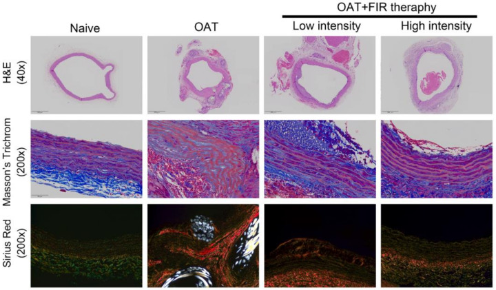
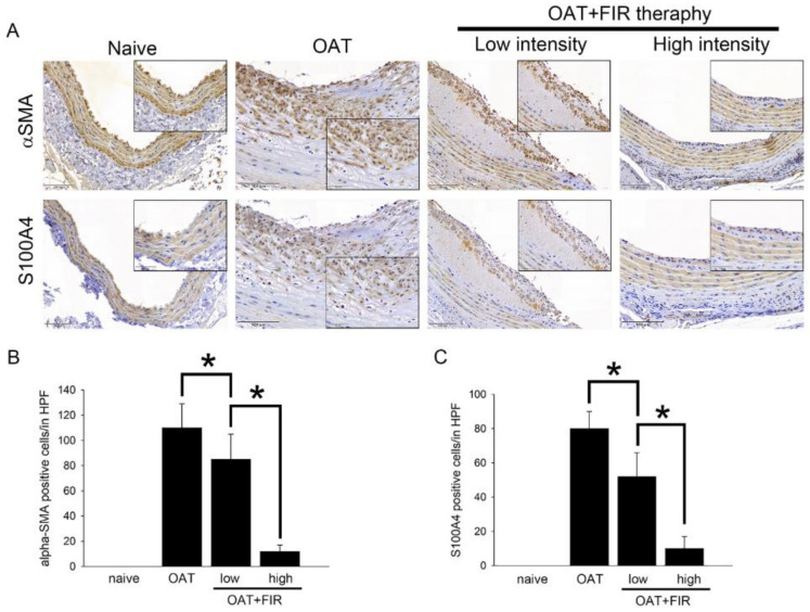
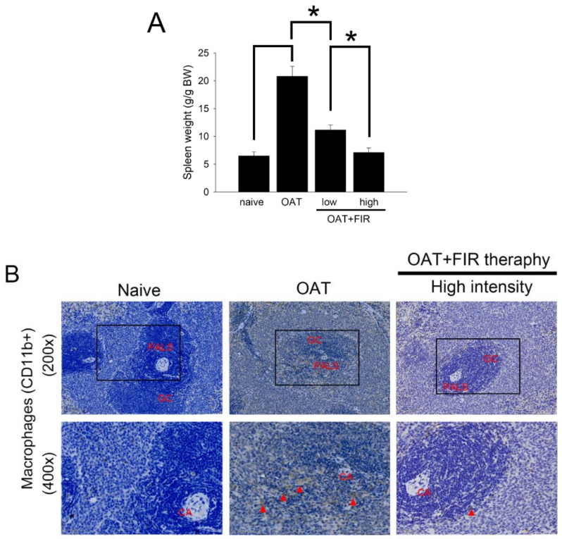
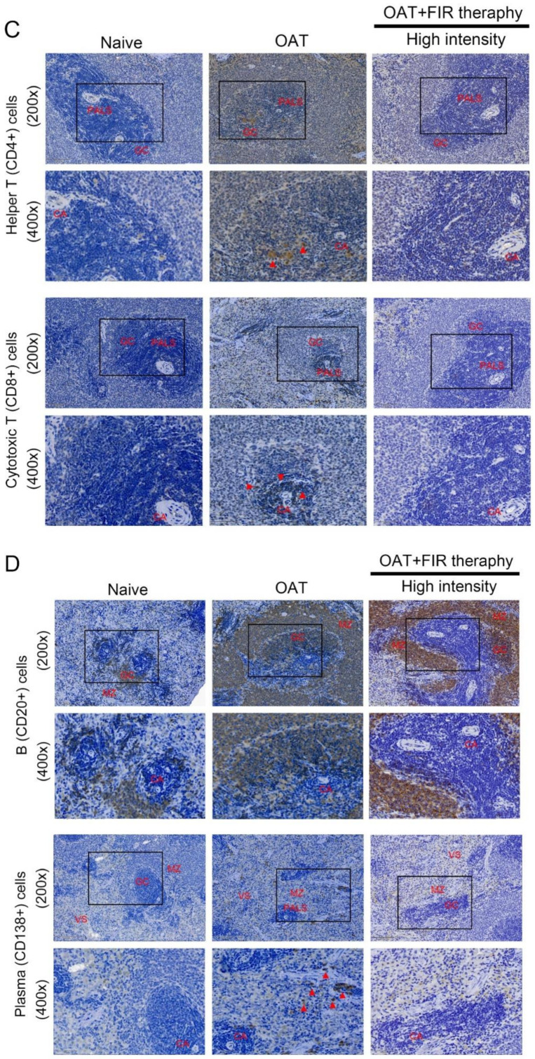
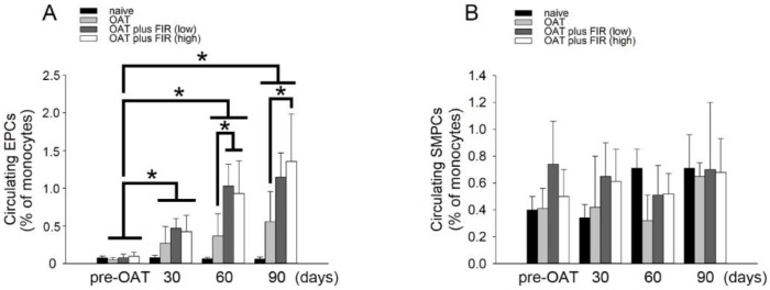
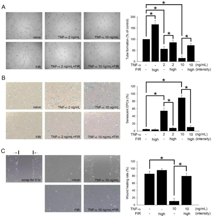
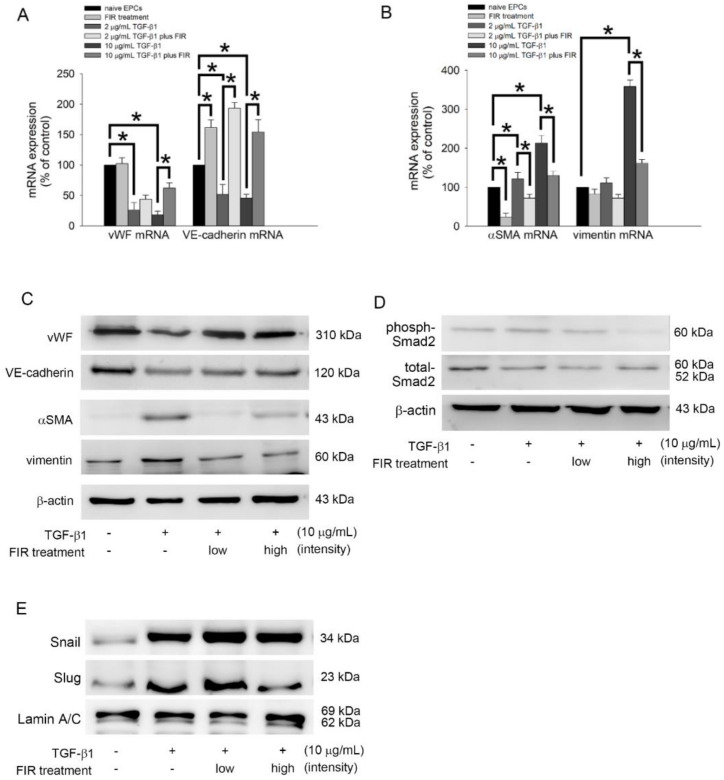
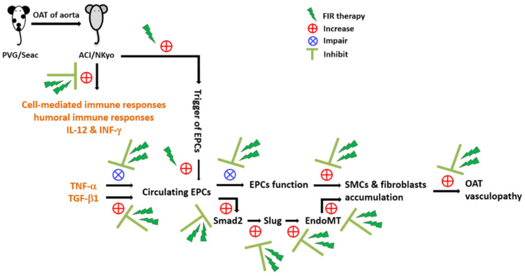
Orthotopic allograft transplantation (OAT) is a major strategy for solid heart and kidney failure. However, the recipient's immunity-induced chronic rejection induces OAT vasculopathy that results in donor organ failure. With the exception of immunosuppressive agents, there are currently no specific means to inhibit the occurrence of OAT vasculopathy. On the other hand, far-infrared (FIR) therapy uses low-power electromagnetic waves given by FIR, with a wavelength of 3-25 μm, to improve human physiological functions. Previous studies have shown that FIR therapy can effectively inhibit inflammation. It has also been widely used in adjuvant therapy for various clinical diseases, especially cardiovascular diseases, in recent years. Thus, we used this study to explore the feasibility of FIR in preventing OAT vasculopathy. In this study, the model of transplantation of an aorta graft from PVG/Seac rat to ACI/NKyo rat, and in vitro model of human endothelial progenitor cells (EPCs) was used. In this report, we presented that FIR therapy decreased the serious of vasculopathy in OAT-recipient ACI/NKyo rats via inhibiting proliferation of smooth muscle cells, accumulation of collagen, and infiltration of fibroblast in the vessel wall; humoral and cell-mediated immune responses were decreased in the spleen. The production of inflammatory proteins/cytokines also decreased in the plasma. Additionally, FIR therapy presented higher mobilization and circulating EPC levels associated with vessel repair in OAT-recipient ACI/NKyo rats. In vitro studies demonstrated that the underlying mechanisms of FIR therapy inhibiting OAT vasculopathy may be associated with the inhibition of the Smad2-Slug axis endothelial mesenchymal transition (EndoMT). Thus, FIR therapy may be the strategy to prevent chronic rejection-induced vasculopathy.

Keywords: endothelial mesenchymal transition; endothelial progenitor cells; far-infrared (FIR) therapy; orthotopic allograft transplantation.


Conflict of interest statement

The authors declare no conflict of interest.

Figures

Figure 1
Figure 1
FIR therapy reduced allograft vasculopathy in OAT-recipient ACI/Nkyo rats. (upper column) Thoracic aortas from donor PVG/Seac rats stained with hematoxylin and eosin. The arrows indicate internal elastic lamina and arrowheads indicate calcified lesions. The images are 40× magnified. (middle column) The integrity of collagen fibers of thoracic aorta cross-sections was observed using Masson’s trichome staining. (lower column) Histopathological features and collagen accumulation of thoracic aorta cross-sections were observed using picrosirius red staining. The slides were observed via light microscopy and polarized light microscopy, respectively (200× magnification).
Figure 2
Figure 2
Administration of FIR therapy is effective against SMC and fibroblast activity in OAT-induced chronic allograft vasculopathy. (A) Immunohistochemistry to assess proliferated SMCs (αSMA) and fibroblasts (S100A4) in rat thoracic aortas from donor PVG/Seac rats. The lumen is uppermost in all sections; the images are 200× magnified. Similar regions are shown as enlarged images (400× magnification) in the black corners. The brown signal indicates αSMA- and S100A4-positive cells. (B,C) The quantification of cells in high power field (HPF) is displayed in (B,C). The graphs demonstrate the accumulation of cells in the aortas of rats. The results are expressed as the mean ± SD. * p < 0.05 was taken into consideration statistically considerable.
Figure 3
Figure 3
FIR therapy decreased splenic T lymphocytes, plasma cells, B lymphocytes, and macrophages activation in the OAT-ACI/NKyo rats. (A) The spleens were dissected from experimental rats after they were sacrificed. The weight of the spleen was analyzed and presented in a bar graph in g/g BW. The results are expressed as the mean ± SD. * p < 0.05 was taken into consideration statistically considerable. (B) Immunohistochemistry was used to analyze the accumulation of splenic CD11b+ macrophages in the OAT-recipient ACI/NKyo rats (CA, central artery; PALS, periarterial lymphatic sheath; GC, germinal center;). The red triangle arrow heads are CD11b+ macrophages. The images in the column are 200× and 400× magnification, respectively. (C) Immunohistochemistry was used to analyze accumulation of splenic CD8+ cytotoxic T cells and CD4+ helper T cells in the recipient rats. The images are presented in 200× and 400× magnification. The CD4+ and CD8+ cells are indicated by red arrow heads. (D) The splenic CD20+ B cells and CD138+ plasma cells accumulation in the OAT-recipient rats (MZ, mantle zone and VS, venous sinuses). The images in the column are 200× and 400× magnification, respectively. The red triangle arrow heads indicate CD138+ cells. The cell nuclei were counted with hematoxylin.
Figure 3
Figure 3
FIR therapy decreased splenic T lymphocytes, plasma cells, B lymphocytes, and macrophages activation in the OAT-ACI/NKyo rats. (A) The spleens were dissected from experimental rats after they were sacrificed. The weight of the spleen was analyzed and presented in a bar graph in g/g BW. The results are expressed as the mean ± SD. * p < 0.05 was taken into consideration statistically considerable. (B) Immunohistochemistry was used to analyze the accumulation of splenic CD11b+ macrophages in the OAT-recipient ACI/NKyo rats (CA, central artery; PALS, periarterial lymphatic sheath; GC, germinal center;). The red triangle arrow heads are CD11b+ macrophages. The images in the column are 200× and 400× magnification, respectively. (C) Immunohistochemistry was used to analyze accumulation of splenic CD8+ cytotoxic T cells and CD4+ helper T cells in the recipient rats. The images are presented in 200× and 400× magnification. The CD4+ and CD8+ cells are indicated by red arrow heads. (D) The splenic CD20+ B cells and CD138+ plasma cells accumulation in the OAT-recipient rats (MZ, mantle zone and VS, venous sinuses). The images in the column are 200× and 400× magnification, respectively. The red triangle arrow heads indicate CD138+ cells. The cell nuclei were counted with hematoxylin.
Figure 4
Figure 4
FIR therapy promotes EPCs mobilization in OAT-recipient ACI/NKyo rats. (A) CD133+/ VEGF+/CD34+ cells (defined as EPCs) mobilization at day 30–90 following OAT in ACI/NKyo rats were analyzed by flow cytometry. (B) CD133+/αSMA+/CD34 cells (defined as SMPCs) mobilization at day 30–90 following OAT in ACI/NKyo rats were studied. Quantification of EPCs (left) and SMPCs (right) in OAT-recipient rats (black bar, naive rats; light gray bar, OAT only rats; dark gray bar, OAT rats with low intensity of FIR therapy; white bar, OAT rats with high intensity of FIR therapy). All results are expressed as the mean ± SD (n = 5). * p < 0.05 was taken into consideration statistically considerable.
Figure 5
Figure 5
FIR treatment promotes the functions of human EPCs. (A) EPCs were stimulated with 2 or 10 ng/mL TNF-α for 24 h with or without high intensity of FIR treatment. An in vitro angiogenesis assay was used to investigate the effect of FIR therapy on EPC neovascularization. Representative photos of in vitro angiogenesis are shown. The graph shows the quantification of tube formation by TNF-α-treated EPCs following FIR treatment. (B) After treating EPCs with TNF-α and high intensity of FIR for 24 h, cell senescence was analyzed; the diagram shows the quantification of senescent EPCs. (C) A migration assay was performed to analyze the effect of FIR on TNF-α-treated EPCs. The 10 ng/mL of TNF-α were treated to EPCs, and adhered to 24 h of FIR treatment before injury scratching. Photos were taken after 8 h of injuring. Counted the migrated EPCs at the denuded location according to the black baseline under 100× high-power field. All data are expressed as the mean ± SD of three independent experiments and as the percentage of the control. * p < 0.05 was taken into consideration statistically considerable.
Figure 6
Figure 6
FIR treatment regulates TGF-β1-induced EndoMT via Smad- and Slug-dependent pathways. (A,B) Human EPCs were exposed to 2 or 10 μg/mL recombinant human TGF-β1 for 5 days with high intensity or without FIR treatment. The α-SMA, VE-cadherin, vWF, and vimentin mRNA expression were evaluated using reverse transcription and qPCR analysis. The expression of related mRNA expression is normalized to the expression of GAPDH mRNA, is presented as a bar graph. All data are expressed as the mean ± SD of five independent experiments and as the percentage of the control. * p < 0.05 was taken into consideration statistically considerable. (C,D) Human EPCs were exposed to 10 μg/mL TGF-β1 for 5 days with low intensity, high intensity or without FIR treatment. The total protein expression of the vWF, VE-cadherin, α-SMA, vimentin, and phosphorylated Smad2 were identified by Western blot analysis. β-actin and total-Smad2 were used as loading controls. (E) Human EPCs were treated with 10 μg/mL TGF-β1 in the presence or absence of FIR treatment for 5 days. Total nuclear lysates were purified, and the levels of Snail and Slug were analyzed using Western blotting; lamin A/C was used as a loading control.
Figure 7
Figure 7
FIR therapy may effectively regulate chronic rejection-induced vasculopathy in OAT rats. Therapy of FIR reduced T and B lymphocytes, plasma cells, and macrophage activation in the spleens of the OAT-recipient ACI/NKyo rats. Lowered the progression of vasculopathy in OAT-recipient ACI/NKyo rats occurred by the inhibition of cell-mediated and humoral immune responses, prevention of cytokines-induced disfunction and EndoMT in EPCs, decrease in collagen damage and pathological accumulation, and proliferation and infiltration of SMCs and fibroblasts in the vessel wall of OAT-recipient ACI/NKyo rats. Therefore, the results highlight the therapeutic roles of FIR and provides a more effective adjuvant therapeutic route in vasculopathy.

References

    1. Sobajima M., Nozawa T., Ihori H., Shida T., Ohori T., Suzuki T., Matsuki A., Yasumura S., Inoue H. Repeated sauna therapy improves myocardial perfusion in patients with chronically occluded coronary artery-related ische-mia. Int. J. Cardiol. 2013;167:237–243. doi: 10.1016/j.ijcard.2011.12.064. - DOI - PubMed
    1. Ohori T., Nozawa T., Ihori H., Shida T., Sobajima M., Matsuki A., Yasumura S., Inoue H. Effect of repeated sauna treatment on exercise tolerance and endothelial function in patients with chronic heart failure. Am. J. Cardiol. 2012;109:100–104. doi: 10.1016/j.amjcard.2011.08.014. - DOI - PubMed
    1. Lin C.C., Chang C.F., Lai M.Y., Chen T.W., Lee P.C., Yang W.C. Far-infrared therapy: A novel treatment to im-prove access blood flow and unassisted patency of arteriovenous fistula in hemodialysis patients. J. Am. Soc. Nephrol. 2007;18:985–992. doi: 10.1681/ASN.2006050534. - DOI - PubMed
    1. Lai C.C., Fang H.C., Mar G.Y., Liou J.C., Tseng C.J., Liu C.P. Post-angioplasty far infrared radiation therapy improves 1-year angioplasty-free hemodialysis access patency of recurrent obstructive lesions. Eur. J. Vasc. Endovasc. Surg. 2013;46:726–732. doi: 10.1016/j.ejvs.2013.09.018. - DOI - PubMed
    1. Tu Y.P., Chen S.C., Liu Y.H., Chen C.F., Hour T.C. Postconditioning with far-infrared irradiation increases heme oxygenase-1 expression and protects against ischemia/reperfusion injury in rat testis. Life Sci. 2013;92:35–41. doi: 10.1016/j.lfs.2012.10.019. - DOI - PubMed