. 2022 May 10;10(5):1106.
doi: 10.3390/biomedicines10051106.

Urinary KIM-1 Correlates with the Subclinical Sequelae of Tubular Damage Persisting after the Apparent Functional Recovery from Intrinsic Acute Kidney Injury

Affiliations

Urinary KIM-1 Correlates with the Subclinical Sequelae of Tubular Damage Persisting after the Apparent Functional Recovery from Intrinsic Acute Kidney Injury

Cristina Cuesta et al. Biomedicines. .

Abstract

Acute kidney injury (AKI) poses an increased risk factor for new AKI episodes, progression to chronic kidney disease, and death. A worsened evolution has been linked to an incomplete renal repair beyond the apparent functional recovery based on plasma creatinine (pCr) normalization. However, structural sequelae pass largely unnoticed due to the absence of specific diagnostic tools. The urinary kidney injury molecule 1 (KIM-1) participates in renal tissue damage and repair and is proposed as a biomarker of early and subclinical AKI. Thus, we study in this paper the evolution of KIM-1 urinary excretion alongside renal tissue sequelae after an intrinsic AKI episode induced by cisplatin in Wistar rats. Creatinine clearance, pCr, proteinuria and the fractional excretion of Na+ and glucose were used to monitor renal function. Renal tissue damage was blindly scored in kidney specimens stained with hematoxylin-eosin and periodic acid-Schiff. KIM-1 urinary excretion and renal mRNA expression were also assessed. Finally, we analyzed urinary KIM-1 in patients apparently recovered from AKI. Our results show that, after the normalization of the standard markers of glomerular filtration and tubular function, the extent of persistent histological findings of tissue repair correlates with the renal expression and urinary level of KIM-1 in rats. In addition, KIM-1 is also elevated in the urine of a significant fraction of patients apparently recovered from an AKI. Besides its potential utility in the early and subclinical diagnosis of renal damage, this study suggests a new application of urinary KIM-1 in the non-invasive follow-up of renal repair after AKI.

Keywords: KIM-1; acute kidney injury; biomarker; subclinical sequelae.


Conflict of interest statement

The authors declare no conflict of interest.

Figures

Figure 1
Figure 1
Flowchart of the study design. B: basal timepoint; D4: day of maximum kidney damage after cisplatin treatment; R0: day of recovery; R1: 1 week after recovery; R2: 2 weeks after recovery; R3: 3 weeks after recovery; R4: 4 weeks after recovery.
Figure 2
Figure 2
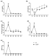
Renal function parameters: plasma creatinine (pCr) (a), creatinine clearance (ClCr) (b), proteinuria (c), fractional excretion of sodium (FENa) (d) and fractional excretion of glucose (FEGlc) (e). B: basa; CDDP: cisplatin treatment (5 mg·kg−1 body weight) group; CT: control group; D4: day of maximum kidney damage after cisplatin treatment; R0: day of recovery; R1: 1 week after recovery; R2: 2 weeks after recovery; R3: 3 weeks after recovery; R4: 4 weeks after recovery. ** p < 0.01 vs. B; ## p < 0.01 vs. CT.
Figure 3
Figure 3
Renal histology. Representative images of kidney specimens stained with haematoxylin and eosin (HE) at different time points of AKI evolution. (B): basal; (D4): day of maximum kidney damage after cisplatin treatment; (R0): day of recovery; (R1): 1 week after recovery; (R2): 2 weeks after recovery; (R3): 3 weeks after recovery; (R4): 4 weeks after recovery. Arrows in D4 indicate widespread detachment of necrotic tubular epithelial cells. Asterisks in R0 and R1 indicate tubular dilatation. Arrows in R2 and R3 indicate intratubular casts.
Figure 4
Figure 4
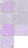
Renal histology. Representative images of kidney specimens stained with periodic acid–Schiff (PAS) at different time points of AKI evolution. (B): basal; (D4): day of maximum kidney damage after cisplatin treatment; (R0): day of recovery; (R1): 1 week after recovery; (R2): 2 weeks after recovery; (R3): 3 weeks after recovery; (R4): 4 weeks after recovery. Arrows in D4 indicate widespread detachment of necrotic tubular epithelial cells. Asterisks in (R0) and (R1) indicate dilated proximal tubules with loss of the brush border. Arrow in R4 indicates an area with interstitial fibrosis and tubular atrophy.
Figure 5
Figure 5
Score of several parameters of histological damage based on haematoxylin–eosin and periodic acid–Schiff staining. B: basal; D4: day of maximum kidney damage after cisplatin treatment; R0: day of recovery; R1: 1 week after recovery; R2: 2 weeks after recovery; R3: 3 weeks after recovery; R4: 4 weeks after recovery. * p < 0.05 vs. B; ** p < 0.01 vs. B.
Figure 6
Figure 6
Urinary excretion of KIM-1 normalized by both urine flow (µg/day) (a) and urine creatinine (ng/mg UCr) (b) and KIM-1 mRNA expression in renal tissue (c). B: basal; CDDP: cisplatin treatment (5 mg·kg−1 body weight) group; CT: control group; D4: day of maximum kidney damage after cisplatin treatment; R0: day of recovery; R1: 1 week after recovery; R2: 2 weeks after recovery; R3: 3 weeks after recovery; R4: 4 weeks after recovery. * p < 0.05 vs. B; # p < 0.05 vs. CT; ** p < 0.01 vs. B; ## p < 0.01 vs. CT.
Figure 7
Figure 7
Urinary excretion of KIM-1 in patients who had recovered from an episode of AKI and controls. (a) Study population characteristics. (b) Urinary KIM-1 level after serum Cr had returned to the values prior to the AKI episode in patients with previous CKD (i.e., chronic) and in patients without previous CKD (i.e., non-chronic), and controls (left panel). Urinary KIM-1 levels (as in B) normalized by urinary creatinine concentration (uCr) (right panel). AU: arbitrary units.

References

    1. Thomas M.E., Blaine C., Dawnay A., Devonald M.A.J., Ftouh S., Laing C., Latchem S., Lewington A., Milford D.V., Ostermann M. The definition of acute kidney injury and its use in practice. Kidney Int. 2015;87:62–73. doi: 10.1038/ki.2014.328. - DOI - PubMed
    1. Ronco C., Bellomo R., Kellum J.A. Acute kidney injury. Lancet. 2019;394:1949–1964. doi: 10.1016/S0140-6736(19)32563-2. - DOI - PubMed
    1. Siew E.D., Davenport A. The growth of acute kidney injury: A rising tide or just closer attention to detail? Kidney Int. 2015;87:46–61. doi: 10.1038/ki.2014.293. - DOI - PMC - PubMed
    1. Macedo E., Garcia-Garcia G., Mehta R.L., Rocco M.V. International Society of Nephrology 0 by 25 Project: Lessons Learned. Ann. Nutr. Metab. 2019;74:45–50. doi: 10.1159/000500345. - DOI - PubMed
    1. Melo F.D.A.F., Macedo E., Bezerra A.C.F., De Melo W.A.L., Mehta R.L., Burdmann E.D.A., Zanetta D.M.T. A systematic review and meta-analysis of acute kidney injury in the intensive care units of developed and developing countries. PLoS ONE. 2020;15:e0226325. doi: 10.1371/journal.pone.0226325. - DOI - PMC - PubMed

LinkOut - more resources