. 2022 May 18;10(5):1160.
doi: 10.3390/biomedicines10051160.

Altered Ocular Surface Health Status and Tear Film Immune Profile Due to Prolonged Daily Mask Wear in Health Care Workers

Affiliations

Altered Ocular Surface Health Status and Tear Film Immune Profile Due to Prolonged Daily Mask Wear in Health Care Workers

Sharon D'Souza et al. Biomedicines. .

Abstract

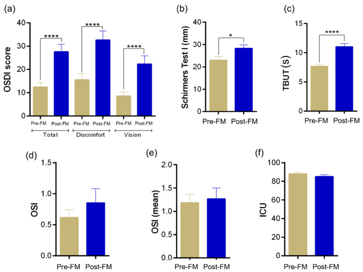
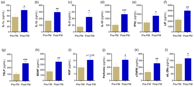
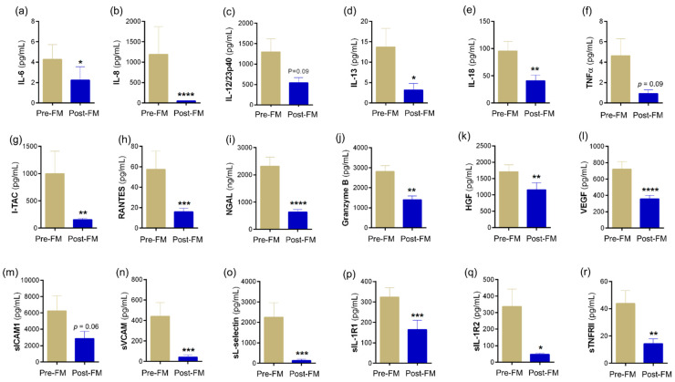
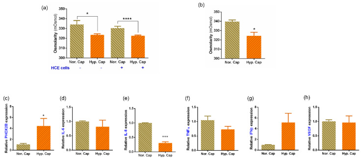
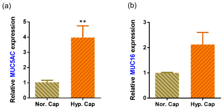
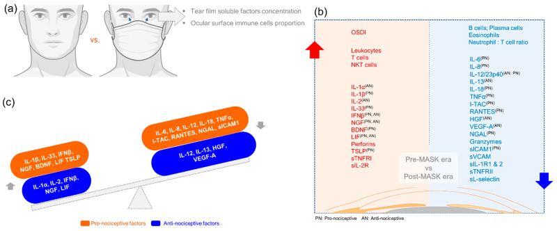
Prolonged daily face mask wearing over several months might affect health of the ocular surface and is reported to be associated with complaints of discomfort and dry-eye-like symptoms. We studied the ocular surface clinical parameters, tear soluble factors and immune cell proportions in ophthalmologists practicing within similar environmental conditions (n = 17) at two time points: pre-face-mask period (Pre-FM; end of 2019) and post-face-mask-wearing period (Post-FM; during 2020 COVID-19 pandemic), with continuous (~8 h/day) mask wear. A significant increase in ocular surface disease index (OSDI) scores without changes in tear breakup time (TBUT), Schirmer's test 1 (ST1) and objective scatter index (OSI) was observed Post-FM. Tear soluble factors (increased-IL-1β, IL-33, IFNβ, NGF, BDNF, LIF and TSLP; decreased-IL-12, IL-13, HGF and VEGF-A) and mucins (MUC5AC) were significantly altered Post-FM. Ex vivo, human donor and corneoscleral explant cultures under elevated CO2 stress revealed that the molecular profile, particularly mucin expression, was similar to the Post-FM tear molecular profile, suggesting hypercapnia is a potential contributor to ocular surface discomfort. Among the immune cell subsets determined from ocular surface wash samples, significantly higher proportions of leukocytes and natural killer T cells were observed in Post-FM compared to Pre-FM. Therefore, it is important to note that the clinical parameters, tear film quality, tear molecular factors and immune cells profile observed in prolonged mask-wear-associated ocular surface discomfort were distinct from dry eye disease or other common ocular surface conditions. These observations are important for differential diagnosis as well as selection of appropriate ocular surface treatment in such subjects.

Keywords: COVID-19; hypercapnia; immune cells; mask; nociception; ocular surface discomfort; soluble factors; tear fluid.


Conflict of interest statement

The authors declare no conflict of interest.

Figures

Figure 1
Figure 1
Status of ocular surface health indices in healthy individuals during the face-mask-wearing era. Bar graphs represent: (a) ocular surface disease index (OSDI) scores and (bf) tear film quality assessments in matched study subjects prior to and during the face-mask-wearing era. (b) Schirmer’s test 1 values in mm/5 min, (c) tear breakup time—TBUT values in s (d) objective scatter index—OSI, (e) mean objective scatter index and (f) lipid layer thickness of the tear film quantified as interferometric colour units—ICU. N = 17; 34 eyes. Pre-FM indicates period where the study subjects were not wearing a face mask (pre COVID-19 era) for an extended period of time. Post-FM indicates period where the same study subjects were wearing a face mask (during the COVID-19 pandemic) for an extended period of time (approximately 8 hours a day). Bar graph indicates mean ± SEM. * p < 0.05, **** p < 0.0001, Wilcoxon matched-pairs signed rank test.
Figure 2
Figure 2
Tear fluid soluble factors increased during face-mask-wearing era. Bar graphs represent the levels of cytokines—(a) IL-1α, (b) IL-1β, (c) IL-2, (d) IL-33, (e) IFNβ, (f) LIF, (g) TSLP; growth factors—(h) BDNF, (i) NGF; cytolytic protein—(j) perforin; and soluble receptors—(k) sTNFRI, (l) sIL-2R α in matched study subjects prior to and during the face-mask-wearing era. IL—interleukin; IFNβ—interferon beta; LIF—leukaemia inhibitory factor, an IL-6 class cytokine; TSLP—thymic stromal lymphopoietin; BDNF—brain-derived neurotrophic factor; NGF—nerve growth factor; sTNFRI—soluble tumour necrosis factor receptor I; sIL-2Rα—soluble form of IL-2 receptor alpha. N = 17; 34 eyes. Pre-FM indicates period where the study subjects were not wearing a face mask (pre COVID-19 era) for an extended period of time. Post-FM indicates the period where the same study subjects were wearing a face mask (during the COVID-19 pandemic) for an extended period of time (approximately 8 hours a day). Bar graph indicates mean ± SEM * p < 0.05, ** p < 0.01, *** p < 0.001; Wilcoxon matched-pairs signed rank test.
Figure 3
Figure 3
Tear fluid soluble factors decreased during face-mask-wearing era. Bar graphs represent the levels of cytokines—(a) IL-6, (c) IL-12/23p40, (d) IL-13, (e) IL-18, (f) TNFα; chemokine—(b) IL-8, (g) I-TAC, (h) RANTES; enzymes—(i) NGAL, (j) granzyme; growth factors—(k) HGF, (l) VEGF; soluble cell adhesion molecules—(m) sICAM1, (n) sVCAM, (o) sL-selectin; soluble receptors—(p) sIL-1R1, (q) sIL-1R2, (r) sTNFRII in matched study subjects prior to and during the face-mask-wearing era. IL—interleukin; TNFα—tumour necrosis factor alpha; I-TAC—interferon-inducible T-cell alpha chemoattractant, CXCL11; RANTES—regulated upon activation, normal T Cell expressed and presumably secreted, CCL5; NGAL—neutrophil gelatinase-associated lipocalin; HGF—hepatocyte growth factor; VEGF—vascular endothelial growth factor-A; sICAM1—soluble intercellular adhesion molecule-1; sVCAM—soluble vascular cell adhesion molecule; sIL-1R1—soluble IL-1 receptor type 1; sIL-1R1—soluble IL-1 receptor type 2; sTNFRII—soluble tumour necrosis factor receptor II; N = 17; 34 eyes. Pre-FM indicates period where the study subjects were not wearing a face mask (pre COVID-19 era) for an extended period of time. Post-FM indicates the period where the same study subjects were wearing a face mask (during the COVID-19 pandemic) for an extended period of time (approximately 8 hours a day). Bar graph indicates mean ± SEM * p < 0.05, ** p < 0.01, *** p < 0.001, **** p < 0.0001; Wilcoxon matched-pairs signed rank test.
Figure 4
Figure 4
Ocular surface immune cell subset proportions during the face-mask-wearing era. Graph indicates percentage of (a) leukocytes—CD45+ cells within the cells analysed in ocular surface wash samples collected from matched study subjects prior to and during the face-mask-wearing era. Graphs indicate the percentages of (b) neutrophils—CD45+CD11b+CD16+CD66b+ cells, (c) monocytes—CD45+CD14+ cells, (d) macrophages—CD45+CD16+ cells, (e) natural killer cells—NK cells—CD45+CD16+CD56+ cells, (f) T cells—CD45+CD3+ cells, (g) NKT cells—CD45+CD3+CD16+CD56+ cells, (h) eosinophils—CD45+CD11b+CD16-CD66b+ cells, (i) B cells—CD45+CD3-CD19+ cells and (j) plasma cells—CD45+CD3-CD19+CD138+ cells within the leukocyte population in ocular surface wash samples collected from matched study subjects prior to and during the face-mask-wearing era. The neutrophil to lymphocyte ratio (k) was also calculated. N = 12; 24 eyes. Pre-FM indicates period where the study subjects were not wearing a face mask (pre COVID-19 era) for an extended period of time. Post-FM indicates period where the same study subjects were wearing a face mask (during the COVID-19 pandemic) for an extended period of time (approximately 8 hours a day). Bar graph indicates mean ± SEM * p < 0.05, *** p < 0.001; Wilcoxon matched-pairs signed rank test.
Figure 5
Figure 5
Effect of hypercapnia on osmolarity and expression of genes in human corneal epithelial monolayer culture and corneoscleral rim explant cultures. (a) Bar graph represents the osmolarity changes in culture media with and without SV40 immortalised human corneal epithelial cells (HCE2) following exposure to either 5% CO2 (normocapnia—Nor. Cap) or 20% CO2 (hypercapnia—Hyp. Cap), in vitro for a period of 24 h. Bar graph represents mean ± SEM of six biological replicates. * p < 0.05, **** p < 0.0001; two-tailed unpaired t-test. (b) Bar graph represents the osmolarity changes in culture media that contained corneoscleral rims following exposure to either 5% CO2 (normocapnia—Nor. Cap) or 20% CO2 (hypercapnia—Hyp. Cap), in vitro for a period of 24 h. Bar graph represents mean ± SEM from three matched biological replicates or corneoscleral rims. * p < 0.05, two-tailed paired sample t-test. (ch) Graphs indicate relative mRNA expression of PHOX2B, IL-6, IL-8, TNFα, IFNβ and VEGF in matched corneoscleral rims following exposure to either 5% CO2 (normocapnia—Nor. Cap) or 20% CO2 (hypercapnia—Hyp. Cap), in vitro for a period of 24 h. The expression of PHOX2B, IL-6, IL-8, TNFα, IFNβ and VEGF was normalised to the expression of β-actin (housekeeping gene). Bar graph indicates mean ± SEM from two technical replicates for each of the three biological replicate experiments. * p < 0.05, *** p < 0.001; two-tailed paired sample t-test. PHOX2B—paired-like homeobox 2B.
Figure 6
Figure 6
Effect of hypercapnia on the expression of mucins in human corneoscleral rim explant cultures. Graphs indicate mean relative mRNA expression of MUC5AC (a) and MUC16 (b) in matched corneoscleral rims following exposure to either 5% CO2 (normocapnia—Nor. Cap) or 20% CO2 (hypercapnia—Hyp. Cap), in vitro for a period of 24 h. The expression of MUC5AC and MUC16 was normalised to expression of β-actin (housekeeping gene). The bar graph indicates mean ± SEM from two technical replicates for each of the three biological replicate experiments. ** p < 0.01; two-tailed paired sample t-test.
Figure 7
Figure 7
Schematic summary of the status of ocular surface clinical indices, proportion of immune cell subsets and tear fluid soluble factor levels during the face-mask-wearing era. Panel (a) is a schematic representation of the study design. Left side (red zone) of panel (b) summarises the list of clinical indices, immune cell subsets and tear soluble factors that increased during the post face mask era compared to the pre face mask era in matched subjects. Right side (blue zone) of panel (b) summarises the list of immune cell subsets and tear soluble factors that decreased during the post face mask era compared to the pre face mask era in matched subjects. Since ocular surface discomfort was one of the main clinical indices that was significantly high, we categorised the tear soluble factors based on their ability to modulate nociception in order to study the relationship between tear soluble factors and the discomfort experienced by the study subjects during the post face-mask-wearing era. Superscript of PN and/or AN beside the various analytes indicates whether they possess pro-nociceptive (PN) or anti-nociceptive (AN) potential as reported in the literature and not limited to ocular surface pain. Panel (c) demonstrates that both pro- and anti-nociceptive factors were dysregulated in tear fluid during post face-mask-wearing era. However, it can be posited that an increase in a select set of pro-nociceptive factors (IL-1β, IL-33, IFNβ, NGF, BDNF, LIF and TSLP) and a decrease in a select set of anti-nociceptive factors (IL-12, IL-13, HGF and VEGF-A) could contribute to ocular surface discomfort without changes in tear fluid dynamics during the post face-mask-wearing period.

References

    1. Zhu N., Zhang D., Wang W., Li X., Yang B., Song J., Zhao X., Huang B., Shi W., Lu R., et al. A Novel Coronavirus from Patients with Pneumonia in China, 2019. N. Engl. J. Med. 2020;382:727–733. doi: 10.1056/NEJMoa2001017. - DOI - PMC - PubMed
    1. Kumar S.U., Kumar D.T., Christopher B.P., Doss C.G.P. The Rise and Impact of COVID-19 in India. Front. Med. 2020;7:250. doi: 10.3389/fmed.2020.00250. - DOI - PMC - PubMed
    1. Center for Systems Science and Engineering (CSSE) at Johns Hopkins University (JHU) COVID-19 Dashboard. 2021. [(accessed on 8 March 2022)]. Available online: https://coronavirus.jhu.edu/map.html.
    1. Shetty R., Lalgudi V.G., Khamar P., Gupta K., Sethu S., Nair A., Honavar S.G., Ghosh A., D’Souza S. Potential ocular and systemic COVID-19 prophylaxis approaches for healthcare professionals. Indian J. Ophthalmol. 2020;68:1349–1356. doi: 10.4103/ijo.IJO_1589_20. - DOI - PMC - PubMed
    1. Desai A.N., Aronoff D.M. Masks and Coronavirus Disease 2019 (COVID-19) JAMA. 2020;323:2103. doi: 10.1001/jama.2020.6437. - DOI - PubMed

LinkOut - more resources