. 2022 May 19;10(5):1171.
doi: 10.3390/biomedicines10051171.

Gasdermin D Deficiency Limits the Transition of Atherosclerotic Plaques to an Inflammatory Phenotype in ApoE Knock-Out Mice

Affiliations

Gasdermin D Deficiency Limits the Transition of Atherosclerotic Plaques to an Inflammatory Phenotype in ApoE Knock-Out Mice

Pauline Puylaert et al. Biomedicines. .

Abstract

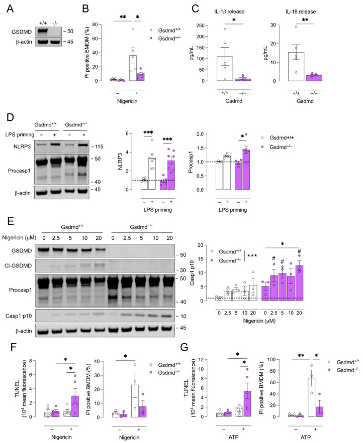
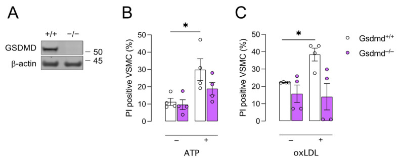
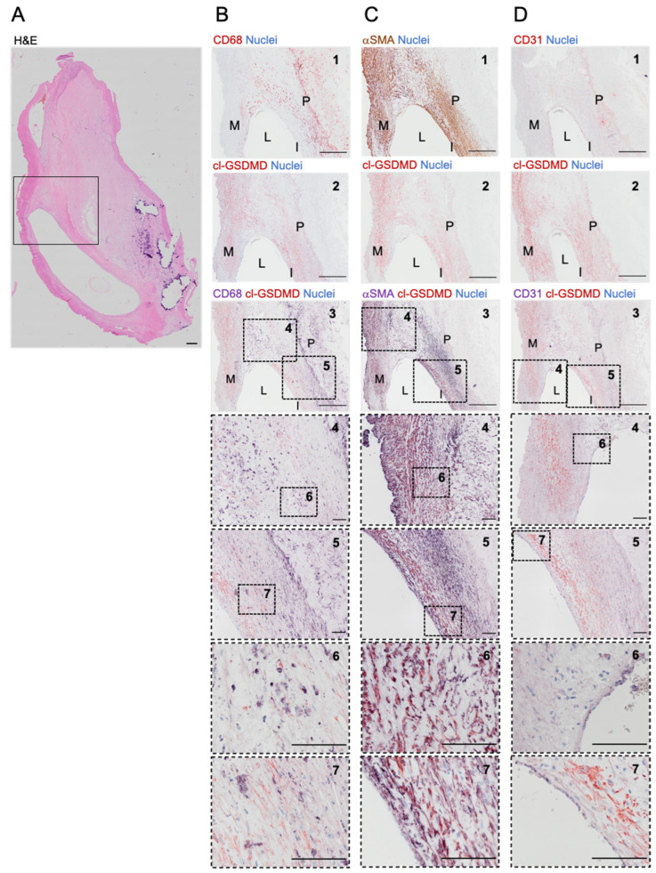
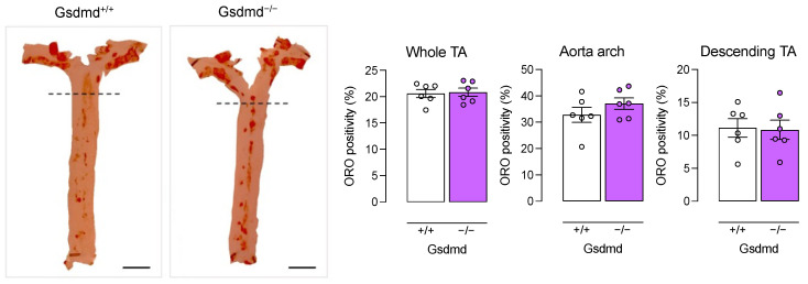
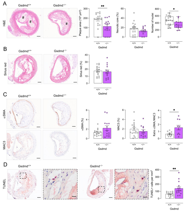
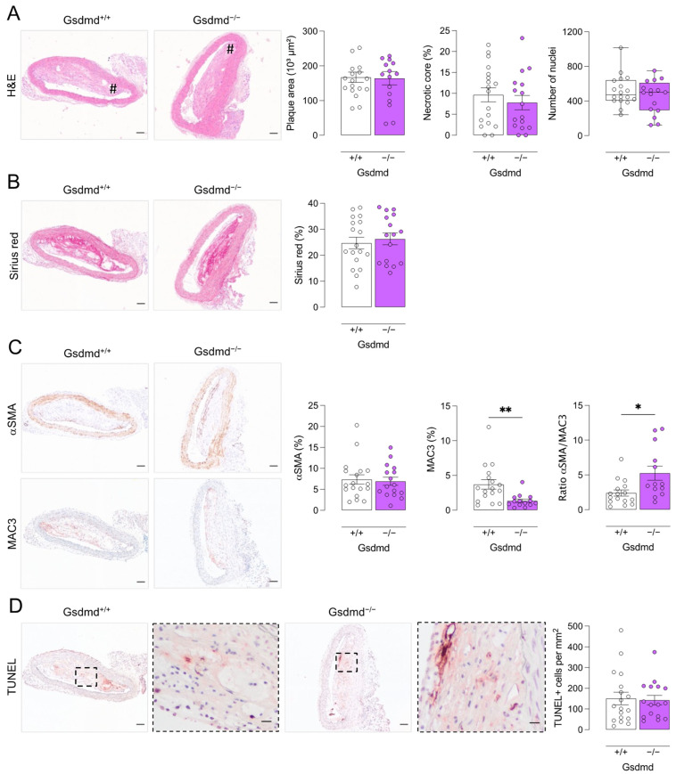
Gasdermin D (GSDMD) is the key executor of pyroptotic cell death. Recent studies suggest that GSDMD-mediated pyroptosis is involved in atherosclerotic plaque destabilization. We report that cleaved GSDMD is expressed in macrophage- and smooth muscle cell-rich areas of human plaques. To determine the effects of GSDMD deficiency on atherogenesis, ApoE-/- Gsdmd-/- (n = 16) and ApoE-/-Gsdmd+/+ (n = 18) mice were fed a western-type diet for 16 weeks. Plaque initiation and formation of stable proximal aortic plaques were not altered. However, plaques in the brachiocephalic artery (representing more advanced lesions compared to aortic plaques) of ApoE-/- Gsdmd-/- mice were significantly smaller (115 ± 18 vs. 186 ± 16 × 103 µm2, p = 0.006) and showed features of increased stability, such as decreased necrotic core area (19 ± 4 vs. 37 ± 7 × 103 µm2, p = 0.03) and increased αSMA/MAC3 ratio (1.6 ± 0.3 vs. 0.7 ± 0.1, p = 0.01), which was also observed in proximal aortic plaques. Interestingly, a significant increase in TUNEL positive cells was observed in brachiocephalic artery plaques from ApoE-/- Gsdmd-/- mice (141 ± 25 vs. 62 ± 8 cells/mm2, p = 0.005), indicating a switch to apoptosis. This switch from pyroptosis to apoptosis was also observed in vitro in Gsdmd-/- macrophages. In conclusion, targeting GSDMD appears to be a promising approach for limiting the transition to an inflammatory, vulnerable plaque phenotype.

Keywords: atherosclerosis; gasdermin D; inflammation; pyroptosis.


Conflict of interest statement

The authors declare no conflict of interest.

Figures

Figure 1
Figure 1
Pyroptosis is inhibited in Gsdmd−/− BMDMs and a switch to apoptosis occurs. Bone marrow-derived macrophages (BMDMs) were isolated from Gsdmd+/+ and Gsdmd−/− mice. (A) Deficiency of GSDMD in Gsdmd−/− BMDMs was confirmed via Western blotting. (BD) Gsdmd−/− and Gsdmd+/+ BMDMs were primed with 100 ng/mL LPS for 4 h followed by treatment with 10 μM nigericin for 2 h. (B) Cell death was measured using propidium iodide (PI) labelling (two-way ANOVA followed by Sidak’s multiple comparison, n = 5 independent experiments) and (C) the release of IL-1β and IL-18 was quantified in the cell supernatant (Mann–Whitney test, n = 5 independent experiments). (D) Western blot analyses of NLRP3 and procaspase (procasp) 1 on lysates of LPS-primed BMDMs (two-way ANOVA followed by Sidak’s multiple comparison, n = 4–7 independent experiments, data are expressed as fold change of target/β-actin ratio). (E) Gsdmd−/− and Gsdmd+/+ BMDMs were primed with 100 ng/mL LPS for 4 h followed by treatment with 2.5–20 μM nigericin for 2 h. Western blot analyses of GSDMD, cleaved (cl)-GSDMD, procaspase 1, and caspase (casp) 1 p10 (two-way ANOVA followed by Dunnett’s multiple comparison between concentrations per genotype, * p < 0.05, *** p < 0.001; two-way ANOVA followed by Sidak’s multiple comparison between genotypes per concentration, # p < 0.05; n = 3 independent experiments, data are expressed as fold change of target/β-actin ratio). (F,G) LPS-primed BMDMs were treated for 1 h with (F) 20 μM nigericin or (G) 5 mM ATP. TUNEL labeling was performed and analyzed via flow cytometry and cell death was measured using PI labelling (two-way ANOVA followed by Sidak’s multiple comparison, n = 3–5 independent experiments). * p < 0.05, ** p < 0.01, *** p < 0.001.
Figure 2
Figure 2
Gsdmd−/− VSMCs are less sensitive to pyroptosis inducers compared to controls. Vascular smooth muscle cells (VSMCs) were isolated from Gsdmd+/+ and Gsdmd−/− mice. (A) Deficiency of GSDMD in Gsdmd−/− VSMCs was confirmed via Western blotting. (B) VSMCs were primed with 50 ng/mL TNFα for 2 h followed by treatment with 5mM ATP for 1 h. (C) VSMCs were treated with 300 μg/mL oxLDL for 48 h. Cell death was measured using PI labelling. * p < 0.05 (two-way ANOVA followed by Sidak’s multiple comparison, n = 4 independent experiments).
Figure 3
Figure 3
Cleaved GSDMD is expressed in human carotid plaques. (A) Overview image of a section from a human carotid artery lesion stained with hematoxylin/eosin. The boxed area corresponds with the region shown in (BD). (BD) Immunohistochemical staining of cleaved (cl)-GSDMD combined with (B) CD68, (C) α-smooth muscle actin (αSMA) or (D) CD31. From top to bottom: 1. Image of section stained for (B) CD68 (red), (C) αSMA (brown), or (D) CD31 (red). 2. Image of section stained for cl-GSDMD (red). 3. Image of double stained section for cl-GSDMD (red) combined with (B) CD68, (C) αSMA, or (D) CD31 (purple). 4–7. Magnifications of dotted frames in images 3–5. Scale bar = 500 μm (A,BD: 1–3), 100 μm (BD: 4–7). Representative images are shown. M = media, L = lumen, I = intima, P = plaque.
Figure 4
Figure 4
Lipid burden in the thoracic aorta is not different between ApoE−/− Gsdmd−/− and ApoE−/− Gsdmd+/+ mice. ApoE−/− Gsdmd−/− and ApoE−/− Gsdmd+/+ mice were fed a WD for 16 weeks. The aortic arch and descending thoracic aorta (TA) were stained en face with Oil Red O to evaluate the plaque burden (Mann–Whitney test, n = 6 mice per group). The dotted line seperates the aortic arch (top) from the descending TA (bottom). Scale bar = 5 mm.
Figure 5
Figure 5
Plaques in the brachiocephalic artery from ApoE−/− Gsdmd−/− mice are smaller but show increased apoptosis. ApoE−/− Gsdmd−/− and ApoE−/− Gsdmd+/+ mice were fed a WD for 16 weeks. Sections of the brachiocephalic artery were stained with (A) hematoxylin/eosin to quantify plaque size, necrotic cores (# hash signs), and cell infiltration; (B) Sirius red to measure total collagen content; (C) anti-MAC3 and anti-α-smooth muscle actin (αSMA) to determine macrophage and smooth muscle cell content, respectively, and to calculate the ratio of αSMA/MAC3 immunoreactivity; (D) TUNEL to count apoptotic cells (dotted boxes are magnified, scale bar = 20 μm). * p < 0.05, ** p < 0.01 (independent samples t-test, boxplot: Mann–Whitney test, n = 10–18 mice per group). Scale bar = 100 μm. Representative images are shown.
Figure 6
Figure 6
Plaques in the proximal aorta from ApoE−/− Gsdmd−/− mice show decreased macrophage infiltration as compared to ApoE−/− Gsdmd+/+ controls. ApoE−/− Gsdmd−/− and ApoE−/− Gsdmd+/+ mice were fed a WD for 16 weeks. Sections of the proximal aorta were stained with (A) hematoxylin/eosin to quantify plaque size, necrotic cores (# hash signs), and cell infiltration; (B) Sirius red to measure total collagen content; (C) anti-MAC3 and anti-α-smooth muscle actin (αSMA) to determine macrophage and smooth muscle cell content, respectively, and to calculate the ratio of αSMA/MAC3 immunoreactivity; (D) TUNEL to count apoptotic cells (dotted boxes are magnified, scale bar = 20 μm). * p < 0.05, ** p < 0.01 (independent samples t-test, boxplot: Mann–Whitney test, n = 13–18 mice per group). Scale bar = 100 μm. Representative images are shown.

Similar articles

Cited by

References

    1. Virmani R., Burke A.P., Farb A., Kolodgie F.D. Pathology of the vulnerable plaque. J. Am. Coll. Cardiol. 2006;47:C13–C18. doi: 10.1016/j.jacc.2005.10.065. - DOI - PubMed
    1. Galluzzi L., Vitale I., Aaronson S.A., Abrams J.M., Adam D., Agostinis P., Alnemri E.S., Altucci L., Amelio I., Andrews D.W., et al. Molecular mechanisms of cell death: Recommendations of the Nomenclature Committee on Cell Death 2018. Cell Death Differ. 2018;25:486–541. doi: 10.1038/s41418-017-0012-4. - DOI - PMC - PubMed
    1. Liu X., Zhang Z., Ruan J., Pan Y., Magupalli V.G., Wu H., Lieberman J. Inflammasome-activated gasdermin D causes pyroptosis by forming membrane pores. Nature. 2016;535:153–158. doi: 10.1038/nature18629. - DOI - PMC - PubMed
    1. Wu D., Chen Y., Sun Y., Gao Q., Yu B., Jiang X., Guo M. Gasdermin family: A promising therapeutic target for cancers and inflammation-driven diseases. J. Cell Commun. Signal. 2020;14:293–301. doi: 10.1007/s12079-020-00564-5. - DOI - PMC - PubMed
    1. Shi J., Zhao Y., Wang K., Shi X., Wang Y., Huang H., Zhuang Y., Cai T., Wang F., Shao F. Cleavage of GSDMD by inflammatory caspases determines pyroptotic cell death. Nature. 2015;526:660–665. doi: 10.1038/nature15514. - DOI - PubMed

LinkOut - more resources