. 2022 May 9;12(5):1180.
doi: 10.3390/diagnostics12051180.

In Vivo Monitoring of Corneal Dendritic Cells in the Subbasal Nerve Plexus during Trastuzumab and Paclitaxel Breast Cancer Therapy-A One-Year Follow-Up

Affiliations

In Vivo Monitoring of Corneal Dendritic Cells in the Subbasal Nerve Plexus during Trastuzumab and Paclitaxel Breast Cancer Therapy-A One-Year Follow-Up

Sebastian Bohn et al. Diagnostics (Basel). .

Abstract

Paclitaxel and trastuzumab have been associated with adverse effects including chemotherapy-induced peripheral neuropathy (CIPN) or ocular complications. In vivo confocal laser scanning microscopy (CLSM) of the cornea could be suitable for assessing side effects since the cornea is susceptible to, i.e., neurotoxic stimuli. The study represents a one-year follow-up of a breast cancer patient including large-area in vivo CLSM of the subbasal nerve plexus (SNP), nerve function testing, and questionnaires during paclitaxel and trastuzumab therapy. Six monitoring sessions (one baseline, four during, and one after therapy) over 58 weeks were carried out. Large-area mosaics of the SNP were generated, and identical regions within all sessions were assigned. While corneal nerve morphology did not cause alterations, the number of dendritic cells (DCs) showed dynamic changes with a local burst at 11 weeks after baseline. Simultaneously, paclitaxel treatment was terminated due to side effects, which, together with DCs, returned to normal levels as the therapy progressed. Longitudinal in vivo CLSM of the SNP could complement routine examinations and be helpful to generate a comprehensive clinical picture. The applied techniques, with corneal structures acting as biomarkers could represent a diagnostic tool for the objective assessment of the severity of adverse events and the outcome.

Keywords: breast cancer; chemotherapy-induced peripheral neuropathy (CIPN); corneal nerves; corneal subbasal nerve plexus (SNP); dendritic cells; identical areas; in vivo large-area confocal laser scanning microscopy (CLSM) of the cornea; one-year follow-up; paclitaxel and trastuzumab.


Conflict of interest statement

The authors declare no conflict of interest.

Figures
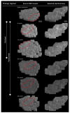
Figure 1
Figure 1
In vivo large-area confocal laser scanning microscopy (CLSM) of the corneal subbasal nerve plexus (SNP) of a breast cancer patient (human epidermal growth factor receptor (HER)-2-positive) at baseline, during, and after paclitaxel and trastuzumab therapy regimen. The detailed patient history and therapy regimen were previously described in [12]. The patient underwent in vivo large-area CLSM at 6 sessions, with a subsequent morphometric assessment of the SNP according to [12]. First column from top to bottom: Timeline of the cancer therapy regimen. The examinations were performed at baseline before treatment, during treatment (at 6 and 11 weeks after baseline examination with combined paclitaxel and trastuzumab therapy; at 44 and 50 weeks after baseline examination with trastuzumab-only therapy), and after treatment (58 weeks after baseline session, 4 weeks after termination of therapy). Paclitaxel therapy was terminated 11 weeks after baseline monitoring due to side effects. Second column from top to bottom: The patient’s SNP before treatment (at baseline), at 6 weeks, at 11 weeks, at 44 weeks, and at 50 weeks (after baseline) during treatment, and at 58 weeks after baseline monitoring (4 weeks after termination of the therapy). The recorded image data were used to generate mosaic SNP images with sizes ranging from 2.5 to 4.2 mm2, analogous to [16]. At baseline and at 6 and 11 weeks after baseline, the same source SNP mosaics were used as in [12]. The assignment of identical areas within all generated mosaics at different sessions was performed in all SNP source mosaics by utilizing so-called k-structures as previously applied in [12], enabling the comparison of the same areas with each other over time (red marked zones). Third column from top to bottom: Enlarged view of identical red marked areas. The size of the identical areas within all mosaics was 0.72 mm2. Nerve fiber morphology remained stable except for minor fluctuations, while dendritic cell numbers showed regional increases at 6 and 11 weeks after the baseline monitoring within selected identical areas.
Figure 2
Figure 2
Total dendritic cell (DC) count within identical corneal areas, nerve function testing score, and questionnaires outcome of a breast cancer patient (human epidermal growth factor receptor (HER)-2-positive) in the course of paclitaxel and trastuzumab breast cancer therapy regimen and monitoring intervals. Total number of dendritic cells: Total DC count within identical areas of the corneal subbasal nerve plexus before therapy at baseline, during therapy (6, 11, 44, and 50 weeks after baseline monitoring), and after therapy at 58 weeks after baseline monitoring. The identical areas throughout all analyzed time points were determined analogous to [12] and accounted for 0.72 mm2. The number of DCs was analyzed manually in triplicate using the ImageJ Cell Counter plugin [17]. At baseline, the DC number was 73 (±3.56), at 6 weeks 167.67 (±8.34), at 11 weeks 472 (±2.94), at 44 weeks 76.33 (±4.1), at 50 weeks 57.33 (±1.25), and at 58 weeks after baseline monitoring number was 65.33 (±2.62). At 6 and 11 weeks, increasing DC numbers were detected, which subsequently decreased to normal levels during the downstream follow-up sessions after cessation of paclitaxel therapy. Neuropathy deficit score (NDS), neuropathy symptom score (NSS), European Organisation for research and Treatment of Cancer Quality of Life Questionnaire-Chemotherapy-Induced Peripheral Neuropathy (EORTC QLQ-CIPN)20, and Functional Assessment of Cancer Therapy (FACT)-Taxane questionnaires: Outcome of nerve function testing and questionnaires during a period of 58 weeks. Nerve function sensitivity was evaluated using NDS and NSS analogous to [13]. While the NSS testing included specific questions regarding neurological symptoms (sensory, motor) with attention to neuropathic pain occurrence and thermoregulatory disturbance, the NDS assessment included measurement of vibration and pain sensation in the feet and legs, monofilament, temperature, and the Achilles tendon reflex [13]. The assessment of the patient’s CIPN symptoms and quality of life (QOL) was analyzed by EORTC QLQ-CIPN20 and FACT-Taxane questionnaires. The EORTC QLQ-CIPN20 is a 20-item questionnaire providing information on CIPN-related symptoms and functional limitations of patients who were exposed to potentially neurotoxic agents with scores ranging from 19 for females and 20 for males (no CIPN-related symptoms) to a maximum of 80 (CIPN related-symptoms) [14]. The FACT-Taxane questionnaire was developed to measure the health-related QOL of patients receiving taxane-containing chemotherapy and is composed of a FACT-general plus a taxane-specific subscale [15]. The FACT-Taxane yields a total score that ranges between 0 and 172, with higher scores reflecting better QOL as well as individual subscale scores such as physical, social, emotional, or functional well-being. [15]. The taxane-specific subscale assesses symptoms related to neurotoxicity, arthralgia, myalgia, and skin discoloration [15]. The NDS was 0 for all monitoring sessions except at 11 weeks after baseline, where it was 1 (no peripheral neuropathy (PNP)). The NSS ranged from 1 to 8, with the highest score at week 11 (baseline: 1—no PNP; 6 weeks: 2—no PNP; 11 weeks: 8—severe PNP; 44 weeks: 5—moderate PNP; 50 weeks: 5—moderate PNP; 58 weeks: 5—moderate PNP). The EORTC QLQ-CIPN20 score ranged between 19 and 24 throughout the whole study period, with the highest level at 11 weeks after baseline monitoring (baseline: 19; 6 weeks: 22; 11 weeks: 24; 44 weeks: 22; 50 weeks: 20; 58 weeks: 23). The FACT-Taxane Total score ranged between 139 and 152, with the highest levels at week 44 (baseline: 150; 6 weeks: 142; 11 weeks: 144; 44 weeks: 152; 50 weeks: 151; 58 weeks: 139). Therapy regimen: Combined paclitaxel and trastuzumab therapy started 4 weeks after baseline monitoring. Paclitaxel treatment was discontinued at 11 weeks after baseline monitoring due to side effects, while trastuzumab was maintained until 54 weeks after baseline. Monitoring interval: A total of six monitoring sessions (one at baseline, four during, and one after therapy) over 58 weeks were carried out. Please note the non-equidistant horizontal time axis.

References

    1. Targeted Cancer Therapies Fact Sheet—National Cancer Institute. [(accessed on 14 March 2022)]; Available online: https://www.cancer.gov/about-cancer/treatment/types/targeted-therapies/t....
    1. Falzone L., Salomone S., Libra M. Evolution of cancer pharmacological treatments at the turn of the third millennium. Front. Pharmacol. 2018;9:1300. doi: 10.3389/fphar.2018.01300. - DOI - PMC - PubMed
    1. Zajaczkowską R., Kocot-Kępska M., Leppert W., Wrzosek A., Mika J., Wordliczek J. Mechanisms of Chemotherapy-Induced Peripheral Neuropathy. Int. J. Mol. Sci. 2019;20:1451. doi: 10.3390/ijms20061451. - DOI - PMC - PubMed
    1. Han Y., Smith M.T. Pathobiology of cancer chemotherapy-induced peripheral neuropathy (CIPN) Front. Pharmacol. 2013;4:156. doi: 10.3389/fphar.2013.00156. - DOI - PMC - PubMed
    1. Breast Cancer. [(accessed on 23 February 2022)]. Available online: https://www.who.int/news-room/fact-sheets/detail/breast-cancer.

LinkOut - more resources