Review
. 2022 May 10;11(10):1598.
doi: 10.3390/cells11101598.

Regulation of Oxidative Phosphorylation of Liver Mitochondria in Sepsis

Affiliations
Review

Regulation of Oxidative Phosphorylation of Liver Mitochondria in Sepsis

Pierre Eyenga et al. Cells. .

Abstract

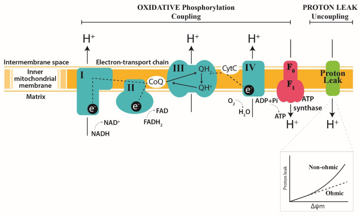
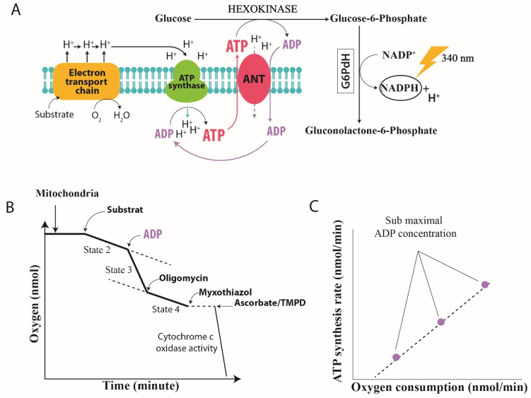
The link between liver dysfunction and decreased mitochondrial oxidative phosphorylation in sepsis has been clearly established in experimental models. Energy transduction is plastic: the efficiency of mitochondrial coupling collapses in the early stage of sepsis but is expected to increase during the recovery phases of sepsis. Among the mechanisms regulating the coupling efficiency of hepatic mitochondria, the slipping reactions at the cytochrome oxidase and ATP synthase seem to be a determining element, whereas other regulatory mechanisms such as those involving proton leakage across the mitochondrial membrane have not yet been formally proven in the context of sepsis. If the dysfunction of hepatic mitochondria is related to impaired cytochrome c oxidase and ATP synthase functions, we need to consider therapeutic avenues to restore their activities for recovery from sepsis. In this review, we discussed previous findings regarding the regulatory mechanism involved in changes in the oxidative phosphorylation of liver mitochondria in sepsis, and propose therapeutic avenues to improve the functions of cytochrome c oxidase and ATP synthase in sepsis.

Keywords: ATP synthase; cytochrome c oxidase; liver; mitochondria; oxidative phosphorylation; sepsis.


Conflict of interest statement

The authors declare no conflict of interest.

Figures

Figure 1
Figure 1
Diagram of the inner mitochondrial membrane, showing three processes that underlie oxidative phosphorylation. (1) The electron transport chain; (2) the uncoupling of oxidation from phosphorylation by leaking H+ across the inner mitochondrial membrane (Leak); and (3) phosphorylation to generate ATP (ATP synthesis). Complex I and II oxidize NADH and FADH2 respectively, transferring the resulting electrons to ubiquinol, which carries electrons to Complex III. Complex III shunts the electrons across the intermembrane space to cytochrome c, which brings electrons to Complex IV. Complex IV then uses the electrons to reduce oxygen to water. The energy liberated by the flow of electrons is used by Complexes I, III and IV to pump protons (H+) out of the mitochondrial matrix into the intermembrane space. This proton gradient generates the mitochondrial membrane potential that is coupled to ATP synthesis by Complex V (ATP synthase) from ADP and inorganic phosphate (Pi). Light green and the insert show the proton leak’s kinetics. The respiration rate increases disproportionately with the membrane potential (non-ohmic proton conductance). NADH, reduced nicotinamide adenine dinucleotide; NAD+, oxidized nicotinamide adenine dinucleotide; FADH, reduced flavin adenine dinucleotide; FAD+, oxidized flavin adenine dinucleotide. The dotted line indicates the electron pathways. The green arrows represent substrate reactions. The black arrows show the proton circuit across the IMM. Complexes I-V are marked as I, II, III and IV in green; ATP synthase is marked in red. Q, ubiquinone; Cyt C, cytochrome c.
Figure 2
Figure 2
Assay chemistry and instrumentation. (A) The ATP:O ratio represents the ratio of ATP synthesis based on oxygen consumption measured simultaneously. It allows us to measure the efficiency of the oxidative phosphorylation. This ratio is determined using the “hexokinase system” based on the regeneration of ADP [17,25], thus making it possible to obtain a stable State 3. The intensity of State 3 can be modulated by increasing concentrations of ADP, resulting in the kinetics of ATP synthesis being obtained as a function of the consumption of oxygen between State 4 (the absence of ATP synthesis) and State 3 (maximum ATP synthesis). The reaction catalyzed by hexokinase (HK) is as follows: synthesized ATP is measured indirectly by evaluating glucose-6-phosphate production during the reaction. Glucose-6-phosphate dehydrogenase (G6PDH) allows the formation of NADPH from glucose-6-phosphate: HK glucose + ATP gGlucose-6-phosphate + ADP. The quantity of NADPH formed during this reaction can then be measured with a spectrophotometer at a wavelength of 340 nm and is directly proportional to the amount of ATP synthesized. (B) Typical trace of mitochondrial respiration. The respiration rate (determined by the slope of the curve) is calculated by the amount of consumed oxygen/minute/mg of protein. The State 2 non-phosphorylating state is respiration in the absence of ATP synthesis. State 3 is respiration in the presence of ATP synthesis. State 4 is the respiration in the presence of oligomycin, an ATP synthase inhibitor. Cytochrome c oxidase activity is the maximal respiration with ascorbate and N,N,N′N′-tetra methyl-p-phenylenediamine (TMPD) as a substrate. (C) ATP:O is the linear relationship between ATP production and oxygen consumption measured with the addition of different amounts of exogenous ADP. The mitochondrial ATP synthesis is initiated by the addition of different amounts of ADP. The intensity of State 3 can be modulated by increasing the concentration of ADP, resulting in the kinetics of ATP synthesis being obtained as a function of oxygen consumption between State 4 (absence of ATP synthesis) and State 3 (maximum ATP synthesis). There is a linear relationship between ATP synthesis and oxygen consumption when the oxidative phosphorylation rate increases by the addition of higher amounts of ADP.
Figure 3
Figure 3
Effect of sepsis on mitochondrial oxidative phosphorylation and compensatory mechanisms for restoring oxidative phosphorylation efficiency (A) Interaction between bacterial lipopolysaccharide (LPS) and toll-like receptors (TLRs) induces pro inflammatory cytokines and triggers radical oxygen species (ROS) and nitric oxide (NO) generation. Increased in ROS and NO production cause damage to ETC components and ATP synthase, and induces a loss of membrane potential through the activation of mitochondrial permeability transition pores (mPTP). Activation of UCP 2 dissipates ROS production but it is not strong enough to prevent the uncoupling of oxidative phosphorylation. (B) Predicted consequences of damage to ETC components and ATP synthase in mitochondrial respiration, and ATP generation and ROS production across the time course of sepsis. (C) Time course of the response of mitochondrial bioenergetic and biogenesis parameters to sepsis, and the expected changes in the coupling efficiency in the recovery phase of sepsis, according to the current knowledge. Arrow (formula image
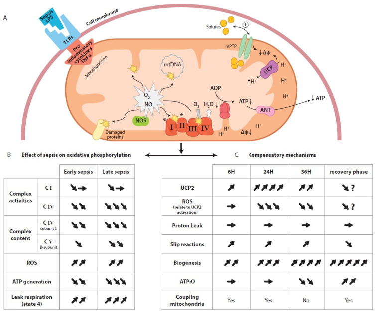
) indicate increase, arrow (formula image) indicate decrease, arrow (formula image) indicate no changes.

References

    1. Angus D.C., Linde-Zwirble W.T., Lidicker J., Clermont G., Carcillo J., Pinsky M.R. Epidemiology of severe sepsis in the United States: Analysis of incidence, outcome, and associated costs of care. Crit. Care Med. 2001;29:1303–1310. doi: 10.1097/00003246-200107000-00002. - DOI - PubMed
    1. Fry D.E., Pearlstein L., Fulton R.L., Polk H.C., Jr. Multiple system organ failure: The role of uncontrolled infection. Arch. Surg. 1980;115:136–140. doi: 10.1001/archsurg.1980.01380020006003. - DOI - PubMed
    1. Nesseler N., Launey Y., Aninat C., White J., Corlu A., Pieper K., Malledant Y., Seguin P. Clinical review: The liver in sepsis. Crit. Care. 2012;16:235. doi: 10.1186/cc11381. - DOI - PMC - PubMed
    1. Wang P., Ba Z.F., Chaudry I.H. Mechanism of hepatocellular dysfunction during early sepsis: Key role of increased gene expression and release of pro inflammatory cytokines tumor necrosis factor and interleukin-6. Arch. Surg. 1991;214:141–148. doi: 10.1001/archsurg.1997.01430280038005. - DOI - PubMed
    1. Wang P., Ba Z.F., Chaudry I.H. Hepatic extraction of indocyanine green is depressed early in sepsis despite increased hepatic blood flow and cardiac output. Arch. Surg. 1991;126:219–224. doi: 10.1001/archsurg.1991.01410260109015. - DOI - PubMed

MeSH terms

Substances