Anatomical Structures Responsible for CTEV Relapse after Ponseti Treatment
- PMID: 35626758
- PMCID: PMC9139296
- DOI: 10.3390/children9050581
Anatomical Structures Responsible for CTEV Relapse after Ponseti Treatment
Abstract
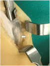
Relapse of deformity after a successful Ponseti treatment remains a problem for the management of clubfoot. An untreated varus heel position and restricted dorsal flexion of the ankle are the main features of recurrences. We analyze the anatomical structures responsible for these recurrences. Materials and methods: During 5 years, 52 children with CTEV (Congenital Talipes Equino Varus) were treated with casts according to the Ponseti method, with a mean number of 7 casts. Closed percutaneous tenotomy was performed in 28 infants. Children were followed monthly and treated with the continuous use of a molded cast. We had 9 children with relapsed clubfeet. During the standing and walking phase, they had a fixed deformity with a varus position of the heel and dorsal flexion of the ankle <10 d. They were surgically treated with the posterolateral approach. Results: In all patients, we found a severe thickening of the paratenon of the Achilles in the medial side, with adhesions with the subcutaneous tissue. The achilles after the previous tenotomy was completely regenerated. The achilles was medially displaced. Conclusions: A severe thickening of the paratenon of the achilles and adhesions with the subcutaneous tissue are anatomical structures in fixed relapsed cases of clubfoot. We treated our patients with an appropriate surgical release.
Keywords: Ponseti method; anatomical structures; club foot; paratenon thickening; relapse club foot.
Conflict of interest statement
The authors declare no conflict of interest.
Figures
References
LinkOut - more resources
Full Text Sources
Miscellaneous


