. 2022 May 6;10(5):855.
doi: 10.3390/healthcare10050855.

Uterine Fibroids and Pregnancy: A Review of the Challenges from a Romanian Tertiary Level Institution

Affiliations

Uterine Fibroids and Pregnancy: A Review of the Challenges from a Romanian Tertiary Level Institution

Mihaela Camelia Tîrnovanu et al. Healthcare (Basel). .

Abstract

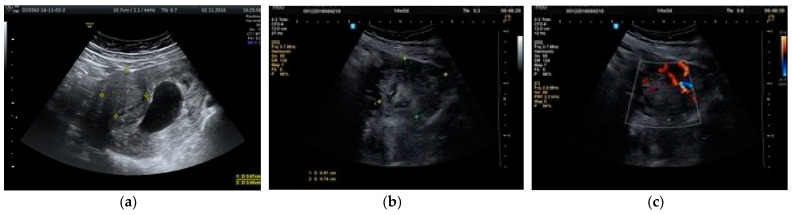
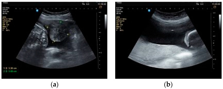
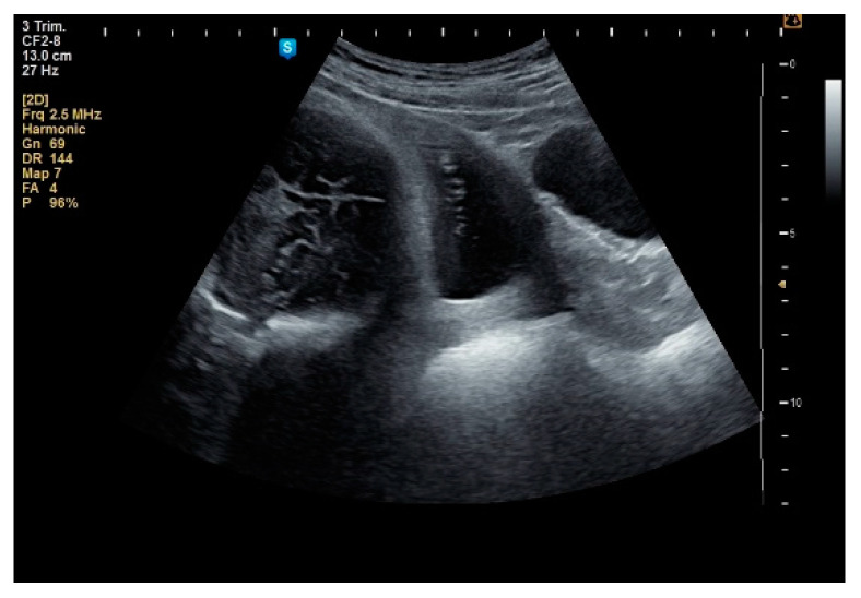
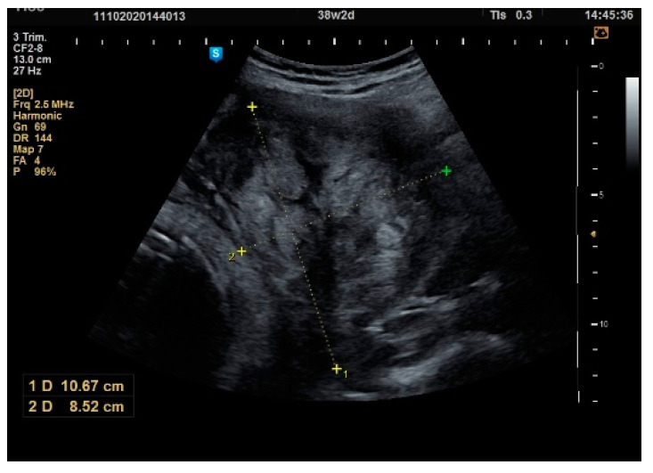
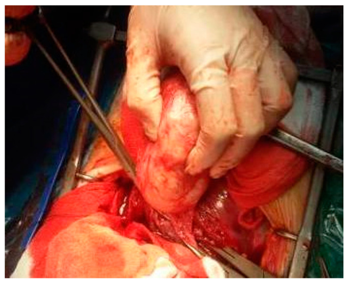
Background and objectives: Uterine fibroids are relatively common in reproductive-age women and are directly linked to pregnancy. There are many debates about performing a myomectomy at the same time as a caesarian section (CS) in such cases due to the risk of a hemorrhage. Our study aims to investigate fibroid features and their evolution in pregnancy, complications of a myomectomy during CS and maternal and fetal obstetric outcomes of pregnancies with fibroids.

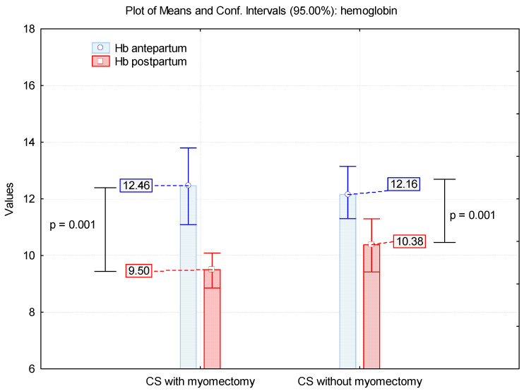
Materials and methods: We realize a prospective study that includes 57 patients with fibroids in pregnancy diagnosed in January 2017-June 2019. We analyze the number, the location and the growth of fibroids during pregnancy and the maternal and fetal outcome. We appreciate the bleeding in patients with a myomectomy and without a myomectomy during CS, using hemoglobin values before and after birth.

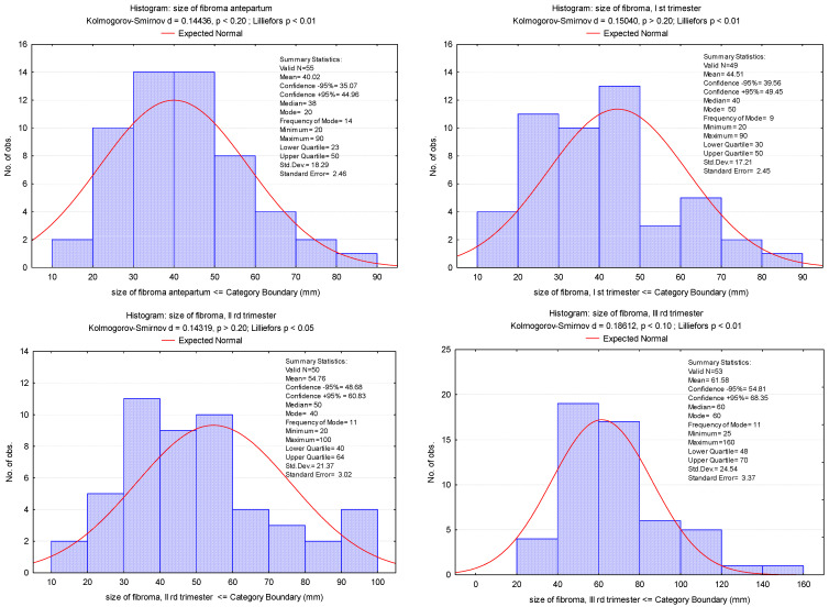
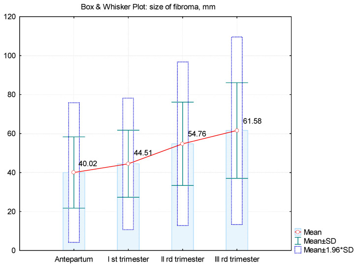
Results: Most of the patients present single fibroids that are 30-160 mm in size, located on the anterior uterine wall. Vaginal delivery is used in 7% of women, whereas 85.96% deliver by CS. In addition, 68% of fibroids are diagnosed in the first trimester. In most cases, the fibroid has maximum growth in the second trimester of pregnancy. The myomectomy rate for fibroids during CS is 24.48. Hemoglobin values showed no statistically significant difference between the two groups with and without myomectomy. The operating time is double for the group with a myomectomy associated with a CS. The results of the obstetric outcomes are abortion in 7% of all patients, whereas premature delivery and births at term are 9.43% and 90.57%, respectively.

Conclusions: The decision of performing a myomectomy during pregnancy can be a challenge and must be performed for selected cases. This procedure may have several benefits, such as avoiding another operation to remove fibroids.

Keywords: myomectomy; obstetric outcome; pregnancy; ultrasound; uterine fibroid.


Conflict of interest statement

The authors declare no conflict of interest.

Figures

Figure 1
Figure 1
Fibroids of small dimensions found during CS (blue arrows), and diagnosis of double uterus (rudimentary left uterus) (yellow arrow).
Figure 2
Figure 2
Uterus with multiple fibroids (blue arrows).
Figure 3
Figure 3
Fibroid dimensions antepartum and in each trimester of pregnancy.
Figure 4
Figure 4
Mean values of dimensions of fibroids increasing during pregnancy.
Figure 5
Figure 5
(a) Ultrasound scan at 9 weeks of pregnancy; fibroid of 36 mm. (b) Ultrasound scan at 14 weeks of pregnancy (same case); fibroid increased to 70 mm. (c) Doppler exam of the same fibroid.
Figure 6
Figure 6
(a). Intracavitary fibroid praevia. (b). Needle inside the amniotic cavity in the superior part of the uterus.
Figure 7
Figure 7
Anterior intramural fibroid and anterior placenta.
Figure 8
Figure 8
Ultrasound scan: pregnancy at term.
Figure 9
Figure 9
Cephalic presentation with intramural fibroid, lateral right, 143/100 mm.
Figure 10
Figure 10
Aspects during CS: (a) After evacuation of the fetus, uterine contractility pushed the fibroid inside the opened uterine cavity; (b) The required myomectomy; (c) Aspect after excision of the fibroid; (d) Suture of the uterine transversal incision.
Figure 11
Figure 11
Subserous fibroid on pregnancy.
Figure 12
Figure 12
Myomectomy during CS.
Figure 13
Figure 13
Uterus after myomectomy.
Figure 14
Figure 14
A 7 cm intracavitary fibroid praevia.
Figure 15
Figure 15
Myomectomy by sectioning the pedicle.
Figure 16
Figure 16
Mean values of hemoglobin in patients with CS with/without myomectomy.

References

    1. Pavone D., Clemenza S., Sorbi F., Fambrini M., Petraglia F. Epidemiology and Risk Factors of Uterine Fibroids. Best Pract. Res. Clin. Obstet. Gynaecol. 2018;46:3–11. doi: 10.1016/j.bpobgyn.2017.09.004. - DOI - PubMed
    1. Parker W.H. Etiology, Symptomatology, and Diagnosis of Uterine Myomas. Fertil. Steril. 2007;87:725–736. doi: 10.1016/j.fertnstert.2007.01.093. - DOI - PubMed
    1. Giuliani E., As-Sanie S., Marsh E.E. Epidemiology and Management of Uterine Fibroids. Int. J. Gynecol. Obstet. 2020;149:3–9. doi: 10.1002/ijgo.13102. - DOI - PubMed
    1. Wang H.-M., Tian Y.-C., Xue Z.-F., Zhang Y., Dai Y.-M. Associations between Uterine Fibroids and Obstetric Outcomes in Twin Pregnancies. Int. J. Gynaecol. Obstet. 2016;135:22–27. doi: 10.1016/j.ijgo.2016.04.013. - DOI - PubMed
    1. Lee H.J., Norwitz E.R., Shaw J. Contemporary Management of Fibroids in Pregnancy. Rev. Obstet. Gynecol. 2010;3:20–27. - PMC - PubMed

LinkOut - more resources