

Early Life Stress Alters Expression of Glucocorticoid Stress Response Genes and Trophic Factor Transcripts in the Rodent Basal Ganglia
- PMID: 35628144
- PMCID: PMC9141219
- DOI: 10.3390/ijms23105333
Early Life Stress Alters Expression of Glucocorticoid Stress Response Genes and Trophic Factor Transcripts in the Rodent Basal Ganglia
Abstract
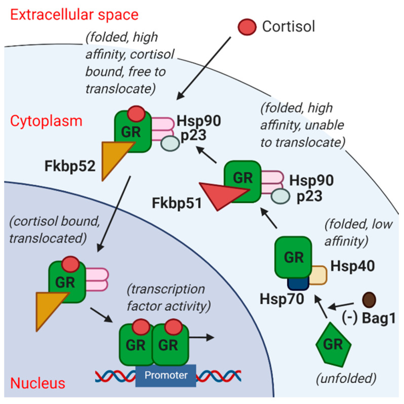
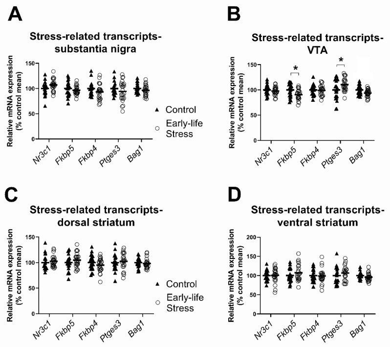
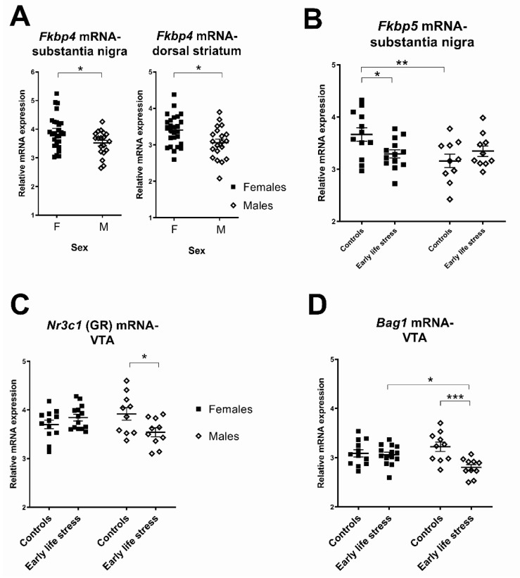
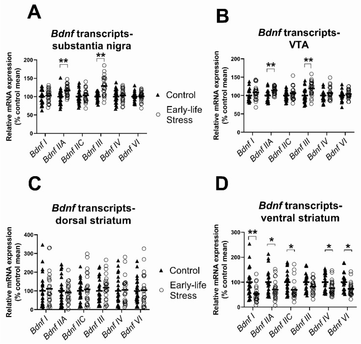
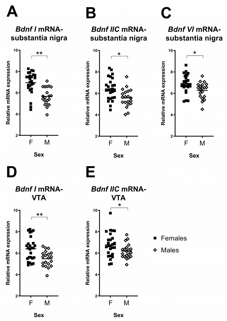
Early life stress shapes the developing brain and increases risk for psychotic disorders. Yet, it is not fully understood how early life stress impacts brain regions in dopaminergic pathways whose dysfunction can contribute to psychosis. Therefore, we investigated gene expression following early life stress in adult brain regions containing dopamine neuron cell bodies (substantia nigra, ventral tegmental area (VTA)) and terminals (dorsal/ventral striatum). Sprague-Dawley rats (14F, 10M) were separated from their mothers from postnatal days (PND) 2-14 for 3 h/day to induce stress, while control rats (12F, 10M) were separated for 15 min/day over the same period. In adulthood (PND98), brain regions were dissected, RNA was isolated and five glucocorticoid signalling-related and six brain-derived neurotrophic factor (Bdnf) mRNAs were assayed by qPCR in four brain regions. In the VTA, levels of glucocorticoid signalling-related transcripts differed in maternally separated rodents compared to controls, with the Fkbp5 transcript significantly lower and Ptges3 transcript significantly higher in stressed offspring. In the VTA and substantia nigra, maternally separated rodents had significantly higher Bdnf IIA and III mRNA levels than controls. By contrast, in the ventral striatum, maternally separated rodents had significantly lower expression of Bdnf I, IIA, IIC, IV and VI transcripts. Sex differences in Nr3c1, Bag1 and Fkbp5 expression in the VTA and substantia nigra were also detected. Our results suggest that early life stress has long-lasting impacts on brain regions involved in dopamine neurotransmission, changing the trophic environment and potentially altering responsiveness to subsequent stressful events in a sex-specific pattern.
Keywords: BDNF; FKBP5; brain-derived neurotrophic factor; dopamine; early life stress; glucocorticoid receptor.
Conflict of interest statement
The authors declare no conflict of interest. The funders had no role in the design of the study; in the collection, analyses, or interpretation of data; in the writing of the manuscript, or in the decision to publish the results.
Figures
Similar articles
-
Short- and long-term effects of intermittent social defeat stress on brain-derived neurotrophic factor expression in mesocorticolimbic brain regions.Neuroscience. 2010 May 19;167(3):598-607. doi: 10.1016/j.neuroscience.2010.02.064. Epub 2010 Mar 3. Neuroscience. 2010. PMID: 20206238 Free PMC article.
-
Overexpression of BDNF in the ventral tegmental area enhances binge cocaine self-administration in rats exposed to repeated social defeat.Neuropharmacology. 2016 Oct;109:121-130. doi: 10.1016/j.neuropharm.2016.04.045. Epub 2016 May 3. Neuropharmacology. 2016. PMID: 27154426 Free PMC article.
-
Dopaminergic neurons in rat ventral midbrain express brain-derived neurotrophic factor and neurotrophin-3 mRNAs.J Comp Neurol. 1994 Apr 15;342(3):321-34. doi: 10.1002/cne.903420302. J Comp Neurol. 1994. PMID: 7912699
-
Neurotrophins in the ventral tegmental area: Role in social stress, mood disorders and drug abuse.Neuroscience. 2014 Dec 12;282:122-38. doi: 10.1016/j.neuroscience.2014.05.028. Epub 2014 May 27. Neuroscience. 2014. PMID: 24875178 Free PMC article. Review.
-
Stress and trauma: BDNF control of dendritic-spine formation and regression.Prog Neurobiol. 2014 Jan;112:80-99. doi: 10.1016/j.pneurobio.2013.10.005. Epub 2013 Nov 6. Prog Neurobiol. 2014. PMID: 24211850 Review.
Cited by
-
Molecular, Pathophysiological, and Clinical Aspects of Corticosteroid-Induced Neuropsychiatric Effects: From Bench to Bedside.Biomedicines. 2024 Sep 19;12(9):2131. doi: 10.3390/biomedicines12092131. Biomedicines. 2024. PMID: 39335644 Free PMC article. Review.
-
Mitigating the impact of adolescence isolation on the development of social anxiety: A potential role for oxytocin.Front Behav Neurosci. 2022 Oct 13;16:1038236. doi: 10.3389/fnbeh.2022.1038236. eCollection 2022. Front Behav Neurosci. 2022. PMID: 36311867 Free PMC article. Review.
-
Effects of Early Stress Exposure on Anxiety-like Behavior and MORC1 Expression in Rats.Biomolecules. 2024 Dec 12;14(12):1587. doi: 10.3390/biom14121587. Biomolecules. 2024. PMID: 39766294 Free PMC article.
-
Sex-dependent preventive effects of prenatal N-acetyl-cysteine on neuronal, emotional and metabolic dysfunctions following exposure to maternal high-fat diet in mice.Transl Psychiatry. 2025 Aug 22;15(1):306. doi: 10.1038/s41398-025-03530-0. Transl Psychiatry. 2025. PMID: 40846701 Free PMC article.
-
Outcomes of early social experiences on glucocorticoid and endocannabinoid systems in the prefrontal cortex of male and female adolescent rats.Front Cell Neurosci. 2023 Dec 20;17:1270195. doi: 10.3389/fncel.2023.1270195. eCollection 2023. Front Cell Neurosci. 2023. PMID: 38174157 Free PMC article.
References
MeSH terms
Substances
Grants and funding
LinkOut - more resources
Full Text Sources
Medical
Miscellaneous
