Review
. 2022 May 12;23(10):5405.
doi: 10.3390/ijms23105405.

Cellulose-Based Nanomaterials Advance Biomedicine: A Review

Affiliations
Review

Cellulose-Based Nanomaterials Advance Biomedicine: A Review

Hani Nasser Abdelhamid et al. Int J Mol Sci. .

Abstract

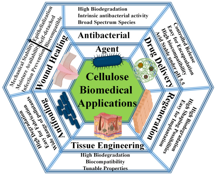
There are various biomaterials, but none fulfills all requirements. Cellulose biopolymers have advanced biomedicine to satisfy high market demand and circumvent many ecological concerns. This review aims to present an overview of cellulose knowledge and technical biomedical applications such as antibacterial agents, antifouling, wound healing, drug delivery, tissue engineering, and bone regeneration. It includes an extensive bibliography of recent research findings from fundamental and applied investigations. Cellulose-based materials are tailorable to obtain suitable chemical, mechanical, and physical properties required for biomedical applications. The chemical structure of cellulose allows modifications and simple conjugation with several materials, including nanoparticles, without tedious efforts. They render the applications cheap, biocompatible, biodegradable, and easy to shape and process.

Keywords: antibacterial; biomedical; cellulose; drug delivery; tissue engineering; wound healing.


Conflict of interest statement

The authors declare no conflict of interest.

Figures

Figure 1
Figure 1
Overview of biomedical applications covered in this review.
Figure 2
Figure 2
Summary of cellulose nanoparticles with sizes and functional groups.
Figure 3
Figure 3
Cellulose-based materials as antibacterial agents.
Figure 4
Figure 4
(a) Synthesis of CNC-Porphyrin; (1) CNC preparation via acid hydrolysis, (2) surface tosylation of CNC, CNC-Tos, (3) synthesis of azide-bearing CNC-N3, (4) click reaction of CNC-N3 with Porphyrin. Reprinted with permission from Ref. [96]. 2011, ACS (2011). (b) Chemical modification of ANCC with Rose Bengal as photosensitizer. Reprinted with permission from Ref. [97]. 2021, American Chemical Society (ACS).
Figure 5
Figure 5
The antibacterial mechanism for porphyrin and quaternary ammonium-modified cellulose under light radiation. Reprinted with permission from Ref. [108]. 2019, John Wiley & Sons.
Figure 6
Figure 6
Schematic representation of suspension and film of MFC and chemical modification with Benzyl Penicillin via esterification. Reprinted with permission from Ref. [129]. 2015, ACS.
Figure 7
Figure 7
Schematic illustration of the immobilization of lysozymes on CNCs for antibacterial activity. Reprinted with permission from Ref. [168]. 2017, ACS.
Figure 8
Figure 8
Wound healing treatment using BC-based dressing: (A) description of the operation on the skin injury model and the dynamic healing of a rat, (B) the progress (0–14 days) of healing for the skin injury model on Wistar rat using gauze and BC-based dressing of two sides, top and bottom (all scale bars equal 10 mm), (C) wound area progression after the injury and (D) wound healing rate. Error bars represent means ± standard deviation (SD) for n = 5 (# p < 0.01).Reprinted with permission from Ref. [220]. 2015, American Chemical Society (ACS, 2015).
Figure 9
Figure 9
(A) Cell migration with and without GO–cellulose nanocomposite; red-dotted lines represent the wound edges, scale bar = 200 μm; (B) in vivo evaluation of the skin wounds of rats with and without GO–cellulose nanocomposite for post-wound induction on days 0, 7, and 21; and (C) the percentage of wound closure: significant differences were evaluated using one-way ANOVA, where *** p < 0.0001. Reprinted with permission from Ref. [230]. 2021, Elsevier.
Figure 10
Figure 10
The synthesis procedure of cellulose-ZIF8 bioink and their processing into the 3D network via 3D printing. Reprinted with permission from Ref. [248]. 2019, John Wiley & Sons.
Figure 11
Figure 11
Schematic representation of the preparation of CNC-SS-PD and their use for gene delivery. Reprinted with permission from Ref. [253]. 2015, ACS.
Figure 12
Figure 12
The 3D printing of NFC–alginate into (A) small grids (7.2 × 7.2 mm2), (B) after squeezing, and (C) restored after squeezing; (DF) 3D-printed human ear in different views. Reprinted with permission from Ref. [280]. 2015, ACS.

References

    1. Quirke V., Gaudillière J.-P. The Era of Biomedicine: Science, Medicine, and Public Health in Britain and France after the Second World War. Med. Hist. 2008;52:441–452. doi: 10.1017/S002572730000017X. - DOI - PMC - PubMed
    1. Wu D., Zhou J., Creyer M.N., Yim W., Chen Z., Messersmith P.B., Jokerst J.V. Phenolic-enabled nanotechnology: Versatile particle engineering for biomedicine. Chem. Soc. Rev. 2021;50:4432–4483. doi: 10.1039/D0CS00908C. - DOI - PMC - PubMed
    1. Nikzamir M., Akbarzadeh A., Panahi Y. An overview on nanoparticles used in biomedicine and their cytotoxicity. J. Drug Deliv. Sci. Technol. 2021;61:102316. doi: 10.1016/j.jddst.2020.102316. - DOI
    1. Liu X., Liang T., Zhang R., Ding Q., Wu S., Li C., Lin Y., Ye Y., Zhong Z., Zhou M. Iron-Based Metal–Organic Frameworks in Drug Delivery and Biomedicine. ACS Appl. Mater. Interfaces. 2021;13:9643–9655. doi: 10.1021/acsami.0c21486. - DOI - PubMed
    1. Swingler S., Gupta A., Gibson H., Kowalczuk M., Heaselgrave W., Radecka I. Recent Advances and Applications of Bacterial Cellulose in Biomedicine. Polymers. 2021;13:412. doi: 10.3390/polym13030412. - DOI - PMC - PubMed

LinkOut - more resources