Review
. 2022 May 12;23(10):5425.
doi: 10.3390/ijms23105425.

Mechanisms of the FMR1 Repeat Instability: How Does the CGG Sequence Expand?

Affiliations
Review

Mechanisms of the FMR1 Repeat Instability: How Does the CGG Sequence Expand?

Elisabetta Tabolacci et al. Int J Mol Sci. .

Abstract

A dynamic mutation in exon 1 of the FMR1 gene causes Fragile X-related Disorders (FXDs), due to the expansion of an unstable CGG repeat sequence. Based on the CGG sequence size, two types of FMR1 alleles are possible: “premutation” (PM, with 56-200 CGGs) and “full mutation” (FM, with >200 triplets). Premutated females are at risk of transmitting a FM allele that, when methylated, epigenetically silences FMR1 and causes Fragile X syndrome (FXS), a very common form of inherited intellectual disability (ID). Expansions events of the CGG sequence are predominant over contractions and are responsible for meiotic and mitotic instability. The CGG repeat usually includes one or more AGG interspersed triplets that influence allele stability and the risk of transmitting FM to children through maternal meiosis. A unique mechanism responsible for repeat instability has not been identified, but several processes are under investigations using cellular and animal models. The formation of unusual secondary DNA structures at the expanded repeats are likely to occur and contribute to the CGG expansion. This review will focus on the current knowledge about CGG repeat instability addressing the CGG sequence expands.

Keywords: CGG repeat; FMR1 gene; dynamic mutations; mechanisms of instability; molecular medicine; neurological disease; repeat expansion disorders.


Conflict of interest statement

The authors declare no conflict of interest.

Figures
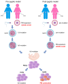
Figure 1
Figure 1
Two models to explain PM-to-FM expansion. The prezygotic model speculates that PM would jump to FM only during maternal germline (meiosis), possibly during recombination, and would further expand during early embryo development (on the left). The postzygotic model hypothesizes that an oocyte carrying a PM would expand to FM during the very first stages of embryogenesis (mitosis) before primordial germ cell (PGC) segregation (on the right). This model does not readily explain why a PM allele transmitted by a father to his daughters cannot expand to FM.
Figure 2
Figure 2
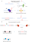
Secondary structures forming at the FRAXA site and their role during DNA synthesis in mitosis (MiDAS). During DNA replication at the FRAXA site, two different secondary structures may form in the CGG repeat sequence: G quadruplex structures on the leading strand and hairpins on the lagging strand. During DNA bidirectional transcription, both nascent RNAs may assume anomalous conformations: the FMR1-mRNA, that is G-rich, is able to form G-quadruplexes, contrariwise the antisense transcript, C-rich, prefers i-motifs. The formation of several secondary structures at the expanded CGG sequence cause failure to complete MiDAS at FRAXA locus. MiDAS starts during S phase of cell cycle with the stalling of DNA polymerase during replication due to the presence of such secondary structures. Endonucleases SLX1/SLX4 induce the formation of a DSB to remove the anomalous structures. The break will be solved during mitosis by the RAD51 recombinase, that binds 3′ end of the cleaved DNA strand and brings it near to the homologous template to complete DNA synthesis via POLD3/Polδ mechanism.
Figure 3
Figure 3
Proposed mechanisms of CGG instability in tissues with low rate of cell division. (A) Oxidation of guanine in DNA strands or during transcription in the displaced DNA strand of the R loop activates BER mechanisms through the recruitment of OGG1 and NEIL1 enzymes, that remove the anomalous base. This damage generates a displacement of the DNA strand favoring hairpin formation. MutL enzymes bind the region containing hairpins in an attempt to remove them; the resulting cleavage produces a DSB which will be repaired, causing the repeat expansion. (B) Large contraction events could be addressed by MMEJ repair mechanism. A DSB may expose sequences of microhomology (MH, in blue) at both sides of the break, which may anneal through the removal of nonhomologous flaps at both ends. Polθ/Polβ complex will fill the gaps, which will be ligated by Lig3 and Lig1, resulting in contraction.
Figure 4
Figure 4
Factors that influence expansion and contraction events during DNA replication at the CGG repeat. The presence of the repeated CGG sequence (blue bars) on the nascent strand induces the formation of hairpins or other secondary structures. At the end of a second round of replication, an expansion (red bars) will result (on the left), whereas the presence of similar secondary structures on the template strand results in a stall with a late restart of the DNA polymerase that causes the exclusion of the repeated tract included in the secondary structure, thus leading to contraction events (green bars) after the second round of replication (on the right). The balance between listed cis and trans elements will hold the repeat sequence stable. An imbalance of these factors will favor instability (contractions or expansions).

References

    1. Pearson C.E., Edamura K.N., Cleary J.D. Repeat instability: Mechanisms of dynamic mutations. Nat. Rev. Genet. 2005;6:729–742. doi: 10.1038/nrg1689. - DOI - PubMed
    1. Depienne C., Mandel J.L. 30 years of repeat expansion disorders: What have we learned and what are the remaining chal-lenges? Am. J. Hum. Genet. 2021;108:764–785. doi: 10.1016/j.ajhg.2021.03.011. - DOI - PMC - PubMed
    1. Verkerk A.J., Pieretti M., Sutcliffe J.S., Fu Y.-H., Kuhl D.P., Pizzuti A., Reiner O., Richards S., Victoria M.F., Zhang F., et al. Identification of a gene (FMR1) containing a CGG repeat coincident with a breakpoint cluster region exhibiting length variation in fragile X syndrome. Cell. 1991;65:905–914. doi: 10.1016/0092-8674(91)90397-H. - DOI - PubMed
    1. Rousseau F., Heitz D., Oberle I., Mandel J.L. Selection in blood cells from female carriers of the fragile X syndrome: Inverse correlation between age and proportion of active X chromosomes carrying the full mutation. J. Med Genet. 1991;28:830–836. doi: 10.1136/jmg.28.12.830. - DOI - PMC - PubMed
    1. Hunter J., Rivero-Arias O., Angelov A., Kim E., Fotheringham I., Leal J. Epidemiology of fragile X syndrome: A systematic review and meta-analysis. Am. J. Med Genet. Part A. 2014;164:1648–1658. doi: 10.1002/ajmg.a.36511. - DOI - PubMed