. 2022 May 13;23(10):5445.
doi: 10.3390/ijms23105445.

Sepsis Encephalopathy Is Partly Mediated by miR370-3p-Induced Mitochondrial Injury but Attenuated by BAM15 in Cecal Ligation and Puncture Sepsis Male Mice

Affiliations

Sepsis Encephalopathy Is Partly Mediated by miR370-3p-Induced Mitochondrial Injury but Attenuated by BAM15 in Cecal Ligation and Puncture Sepsis Male Mice

Pratsanee Hiengrach et al. Int J Mol Sci. .

Abstract

BAM15 (a mitochondrial uncoupling agent) was tested on cecal ligation and puncture (CLP) sepsis mice with in vitro experiments. BAM15 attenuated sepsis as indicated by survival, organ histology (kidneys and livers), spleen apoptosis (activated caspase 3), brain injury (SHIRPA score, serum s100β, serum miR370-3p, brain miR370-3p, brain TNF-α, and apoptosis), systemic inflammation (cytokines, cell-free DNA, endotoxemia, and bacteremia), and blood-brain barrier (BBB) damage (Evan's blue dye and the presence of green fluorescent E. coli in brain after an oral administration). In parallel, brain miR arrays demonstrated miR370-3p at 24 h but not 120 h post-CLP, which was correlated with metabolic pathways. Either lipopolysaccharide (LPS) or TNF-α upregulated miR370-3p in PC12 (neuron cells). An activation by sepsis factors (LPS, TNF-α, or miR370-3p transfection) damaged mitochondria (fluorescent color staining) and reduced cell ATP, possibly through profound mitochondrial activity (extracellular flux analysis) that was attenuated by BAM15. In bone-marrow-derived macrophages, LPS caused mitochondrial injury, decreased cell ATP, enhanced glycolysis activity (extracellular flux analysis), and induced pro-inflammatory macrophages (iNOS and IL-1β) which were neutralized by BAM15. In conclusion, BAM15 attenuated sepsis through decreased mitochondrial damage, reduced neuronal miR370-3p upregulation, and induced anti-inflammatory macrophages. BAM15 is proposed to be used as an adjuvant therapy against sepsis hyperinflammation.

Keywords: BAM15; cecal ligation and puncture; extracellular flux; sepsis; uncoupling agent.


Conflict of interest statement

The authors declare no conflict of interest.

Figures

Figure 1
Figure 1
Characteristics of cecal ligation and puncture (CLP) sepsis or sham mice at 24 h after surgery with BAM15 or vehicle (control) as evaluated by survival analysis (n = 20/group) (A), total white blood cell (WBC) and neutrophils in peripheral blood (B,C), renal function (blood urea nitrogen, serum creatinine, and kidney histological score) (DF), liver injury (alanine transaminase and liver histological score) (G,H), spleen apoptosis (activated caspase 3) (I), encephalopathy clinical score (SHIRPA score; see Methods) (J), and encephalopathy parameters in serum (S100β and miR370-3p) (K,L), brain miR370-3p, brain TNF-α, and brain apoptosis (activated caspase 3) (MO) are shown (n = 6–8/group). #, p < 0.05 vs. sham; * p < 0.05 vs. the indicated groups as determined by ANOVA with Tukey’s analysis. The survival analysis is calculated by Log-rank test. Data from sham mice with BAM15 are not demonstrated due to the non-significant difference to the sham group.
Figure 2
Figure 2
Representative pictures of Hematoxylin and Eosin (H&E) staining (kidneys and livers) (A,B) and activated caspase 3 immunohistochemistry (apoptosis) (spleen and brain) (C,D) of cecal ligation and puncture (CLP) sepsis or sham mice at 24 h after surgery with BAM15 or vehicle (control) are demonstrated. The pictures from sham mice with BAM15 are not shown due to the non-significant difference to the sham group.
Figure 3
Figure 3
Characteristics of cecal ligation and puncture (CLP) sepsis or sham mice at 24 h after surgery with BAM15 or vehicle (control) as evaluated by serum cytokines (TNF-α, IL-6, and IL-10) (AC), serum cell-free DNA (cf-DNA) (D), serum endotoxin (E), and bacteremia (F) are demonstrated (n = 6–8/group). #, p < 0.05 vs. sham; *, p < 0.05 vs. the indicated groups as determined by ANOVA with Tukey’s analysis. Data from sham mice with BAM15 are not demonstrated due to the non-significant difference to the sham group.
Figure 4
Figure 4
The abnormal permeability in the brain (Evan’s blue dye assay) (A) and abundance (fluorescent intensity) of green-fluorescent-producing (GFP) E. coli in the mouse brains at 24 h post-surgery with oral GFP-E. coli administration at 6 h prior to the operation (sham or CLP) with the representative fluorescent figures (B,C) are demonstrated (n = 6–8/group). #, p < 0.05 vs. Sham; *, p < 0.05 vs. the indicated groups as determined by ANOVA with Tukey’s analysis. Data from sham mice with BAM15 are not demonstrated due to the non-significant difference to the sham group.
Figure 5
Figure 5
Heatmap illustration of microRNA arrays from the brains of mice with 24 h post-sham (sham), 24 h post-cecal ligation and puncture (CLP), and 120 h post-CLP (120 h post-sham group is not performed due to the similarity to 24 h post-sham) (A) (n = 3/group) and the predicted pathways that are associated with miR370-3p from the Reactome database are demonstrated (B). The arrowhead indicates miR370-3p on the analysis. The letters in red color are the predicted genes that are associated with miR370-3p from the Reactome analysis (more details on Figure 6) (This picture was provided by Biorender.com (accessed on 5 January 2022)).
Figure 6
Figure 6
The figure shows binding energy of miR370-3p to the possible target in the metabolic pathway (Reactome analysis).
Figure 7
Figure 7
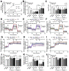
The expression of miR370-3p in neuron cells (PC-12 cell line) after stimulation with lipopolysaccharide (LPS) and TNF-α (A) and characteristics of PC-12 cells after 48 h stimulation by LPS, miR370-3p transfection, or TNF-α with or without BAM15, in comparison with control media (media) or BAM15 alone, as evaluated by mitochondrial membrane potential (B), total cell ATP (C), cell energy status through mitochondria (oxygen consumption rate; OCR) and glycolysis (extracellular acidification rate; ECAR) with several parameters (maximal respiration, respiratory reserve, area under the curve (AUC) of ECAR) (DL) are also demonstrated. #, p < 0.05 vs. control; *, p < 0.05 vs. the indicated groups. The statistical analysis was determined by repeated measure ANOVA (time-point data) and ANOVA with Tukey’s method. #, p < 0.05 vs. control; independent triplicate experiments were performed. Notably, media and BAM15 control groups of miR370-3p transfection were also transfected by miR370-3p mimic (miR-negative control). MFI, mean fluorescent intensity.
Figure 8
Figure 8
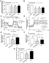
Characteristics of bone-marrow-derived macrophages after 24 h stimulation by LPS, with or without BAM15, in comparison with control media (media), as evaluated by cell energy status through mitochondrial membrane potential (A), total cell ATP (B), and extracellular flux analysis using mitochondria (oxygen consumption rate; OCR) and glycolysis (extracellular acidification rate; ECAR) with several parameters (basal respiration, maximal respiration, respiratory reserve, and area under the curve (AUC) of ECAR) (CG) are demonstrated. The data from macrophages with BAM15 without LPS are not demonstrated in (CG) due to the non-difference from the control group. #, p < 0.05 vs. control; *, p < 0.05 vs. the indicated groups as determined by ANOVA with Tukey’s analysis. #, p < 0.05 vs. control; independent triplicate experiments were performed. MFI, mean fluorescent intensity.
Figure 9
Figure 9
Characteristics of bone-marrow-derived macrophages after 24 h stimulation by LPS, with or without BAM15, in comparison with control media (media), as evaluated by gene expression of inflammatory cytokines (TNF-α, IL-6, and IL-10) (AC), M1 macrophage polarization (pro-inflammation) (iNOS and IL-1β) (D,E), and M2 macrophage polarization (anti-inflammation) (Arginase-1, Fizz-1, and TGF-β) (FH) are demonstrated. The data from macrophages with BAM15 without LPS are not demonstrated due to the non-difference from control group. #, p < 0.05 vs. control; *, p < 0.05 vs. the indicated groups as determined by ANOVA with Tukey’s analysis. #, p < 0.05 vs. control; independent triplicate experiments were performed.
Figure 10
Figure 10
The proposed working hypothesis demonstrates the impacts of bacterial sepsis on brain cells and macrophages. In the brain, blood–brain barrier (BBB) defects in sepsis facilitate the translocation of several molecules, including cytokines, lipopolysaccharide (LPS), and bacteria into the brain. LPS and cytokines (especially TNF-α) upregulated miR370-3p through TLR-4 and TNF receptor (TNFR), respectively, that vigorously activate mitochondria leading to mitochondrial injury. In macrophages, several molecules in the blood during sepsis also activate macrophages, causing hyper-inflammatory responses. However, BAM15 blocks mitochondrial over-activity, which is beneficial for both brain cells and macrophages, resulting in less severe sepsis and sepsis encephalopathy.

Similar articles

Cited by

References

    1. Amornphimoltham P., Yuen P.S.T., Star R.A., Leelahavanichkul A. Gut Leakage of Fungal-Derived Inflammatory Mediators: Part of a Gut-Liver-Kidney Axis in Bacterial Sepsis. Dig. Dis. Sci. 2019;64:2416–2428. doi: 10.1007/s10620-019-05581-y. - DOI - PubMed
    1. Doi K., Leelahavanichkul A., Yuen P.S., Star R.A. Animal models of sepsis and sepsis-induced kidney injury. J. Clin. Investig. 2009;119:2868–2878. doi: 10.1172/JCI39421. - DOI - PMC - PubMed
    1. Chancharoenthana W., Leelahavanichkul A. Acute kidney injury spectrum in patients with chronic liver disease: Where do we stand? World J. Gastroenterol. 2019;25:3684–3703. doi: 10.3748/wjg.v25.i28.3684. - DOI - PMC - PubMed
    1. Wang D., Yin Y., Yao Y. Advances in sepsis-associated liver dysfunction. Burn. Trauma. 2014;2:97–105. doi: 10.4103/2321-3868.132689. - DOI - PMC - PubMed
    1. Manrique-Caballero C.L., Del Rio-Pertuz G., Gomez H. Sepsis-Associated Acute Kidney Injury. Crit. Care Clin. 2021;37:279–301. doi: 10.1016/j.ccc.2020.11.010. - DOI - PMC - PubMed