. 2022 May 13;23(10):5470.
doi: 10.3390/ijms23105470.

Non-Apoptotic Caspase-3 Activation Mediates Early Synaptic Dysfunction of Indirect Pathway Neurons in the Parkinsonian Striatum

Affiliations

Non-Apoptotic Caspase-3 Activation Mediates Early Synaptic Dysfunction of Indirect Pathway Neurons in the Parkinsonian Striatum

Tim Fieblinger et al. Int J Mol Sci. .

Abstract

Non-apoptotic caspase-3 activation is critically involved in dendritic spine loss and synaptic dysfunction in Alzheimer's disease. It is, however, not known whether caspase-3 plays similar roles in other pathologies. Using a mouse model of clinically manifest Parkinson's disease, we provide the first evidence that caspase-3 is transiently activated in the striatum shortly after the degeneration of nigrostriatal dopaminergic projections. This caspase-3 activation concurs with a rapid loss of dendritic spines and deficits in synaptic long-term depression (LTD) in striatal projection neurons forming the indirect pathway. Interestingly, systemic treatment with a caspase inhibitor prevents both the spine pruning and the deficit of indirect pathway LTD without interfering with the ongoing dopaminergic degeneration. Taken together, our data identify transient and non-apoptotic caspase activation as a critical event in the early plastic changes of indirect pathway neurons following dopamine denervation.

Keywords: Parkinson’s disease; Q-VD-OPh; caspase-3; dendritic spines; long-term depression; mice; spiny projection neurons; striatum.


Conflict of interest statement

The authors declare no conflict of interest.

Figures
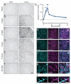
Figure 1
Figure 1
Caspase-3 is transiently upregulated in the DA-denervated striatum. (AF) Striatal sections immunostained with a caspase-3 antibody depict the time course of caspase-3 activation in the DA-denervated striatum (right) relative to the control side (left) post 6-OHDA lesion. Scale bar: 100 μm. (G) Densitometric analysis reveals an increase of caspase-3 levels in the DA-denervated striatum at three- and five-days post-lesion as compared to sham-lesioned animals (represented by “Day 0” datapoint). * p < 0.05, ** p < 0.01 vs. Sham, ANOVA and post hoc Bonferroni test, N = 4–7. (HK) Dual-antigen immunostaining demonstrates caspase-3 co-labeling with markers for astrocytes (GFAP and S100β; H,I), microglia (IBA1; J) and iSPN dendrites (eGFP; K). The latter were labeled using an AAV-based strategy (see methods). (L) Zoom-in of inset in (K) shows puncta of caspase-3 expression within iSPN dendrites. Scale bar: 20 µm (HJ), 10 μm (K), 3 µm (L).
Figure 2
Figure 2
The peak of caspase-3 activation follows the loss of striatal DAergic innervation. (A) Western blot analysis of striatal TH levels shows an exponential decline of DAergic innervation between one and five days after the 6-OHDA lesion (dotted line depicts a one-phase decay fit with the given R² value). **** p < 0.0001 vs. one day post-lesion, ANOVA and post hoc Bonferroni test, N = 6–7. Right: Representative immunoblots with TH bands (60 kDa). (B) Caspase-3 activation was assessed as a ratio of the cleaved (19 and 17 kDa bands) over the uncleaved protein (35 kDa). The ratios are shown relative to the corresponding intact striatum and confirm a peak of caspase-3 activation at five days post-lesion. * p < 0.05 vs. one day post-lesion, ANOVA and post hoc Bonferroni test, N = 6–7. Right: Example of caspase-3-stained blots. (C,D) TUNEL staining was used to reveal apoptotic nuclei in sections through the caudal diencephalon (encompassing the MFB, C) and DA-denervated striatum (D). At the peak of caspase activation (five days post-6-OHDA lesion), no TUNEL positivity was found in the striatum (D,D’), whereas numerous cells displayed apoptotic features along the track of the 6-OHDA infusion (C,C’). (C’,D’) High magnification of (C,D), as indicated. TUNEL (magenta) is overlaid with TO-PRO-3 counterstain of neuronal nuclei (gray). Among the TUNEL-positive cells, some are colabeled with TO-PRO-3 and others are not, indicating different stages of apoptosis. Scale bars: 1 mm (C), 40 µm (C’) 10 µm (D’).
Figure 3
Figure 3
Early synaptic deficits after DA denervation are prevented by pharmacological caspase inhibition. (A) Dendritic spine density of iSPNs is reduced 5–6 days post-6-OHDA lesion. This loss is prevented by systemic treatment with the caspase inhibitor Q-VD-OPh (“QVD”). **** p < 0.0001 vs. 6-OHDA + vehicle, ANOVA and post hoc Bonferroni tests. N = 7–13 cells. (B) Two-photon images of iSPN dendrites, visualized after dye-filling the neurons through the patch pipette. Maximum-intensity projections of dendrites from controls and 6-OHDA-lesioned mice treated with vehicle or QVD are shown from left to right. Scale bar: 5 μm. (C) Sketch of the LTD recording paradigm. GFP-positive iSPNs were patched in the dorsolateral striatum and the stimulus electrode was placed near the border of the cortex. To induce LTD, high-frequent input stimulation (1 s at 100 Hz) was paired with postsynaptic depolarization. (D) Corticostriatal EPSCs are depressed using this protocol in control iSPNs (black). However, EPSC amplitudes are unchanged after HFS in 6-OHDA lesioned mice at 5–6 days after lesion (magenta). Co-treatment with QVD rescues this deficit, and LTD is readily induced (cyan). (E) Example traces from the recordings. Single EPSCs are shown before (black) and after (red) HFS. (F) Quantification shows loss and rescue of HFS-LTD. *** p < 0.001, **** p < 0.0001 vs. 6-OHDA + vehicle, ANOVA and post hoc Bonferroni test, N = 6-8 cells (G) After HFS-LTD induction, paired-pulse ratios (PPRs) are increased in control iSPNs (black) and unchanged in the DA-denervated striatum (magenta). Treatment with QVD rescues the LTD induction, with concomitant increase in PPRs (cyan). * p < 0.05, paired t-test, N = 6-8 cells.
Figure 4
Figure 4
Q-VD-OPh treatment does not protect against 6-OHDA-induced dopaminergic degeneration. (A,B) Low-magnification images of striatal (A) and nigral (B) sections immunostained for TH, with the side ipsilateral to the 6-OHDA lesion on the right. Scale bar: 400 μm (C) Densitometric analysis shows that the loss of striatal DAergic innervation does not differ between vehicle- and QVD-treated animals. **** p < 0.0001 vs. sham, ANOVA and post hoc Bonferroni test, N = 4–5. (D) Stereological cell counts show that the 6-OHDA-induced loss of nigral DA neurons is not prevented by QVD treatment. * p < 0.05 vs. sham, ANOVA and post hoc Bonferroni test, N = 4–5.

References

    1. Li J., Yuan J. Caspases in apoptosis and beyond. Oncogene. 2008;27:6194–6206. doi: 10.1038/onc.2008.297. - DOI - PubMed
    1. Riedl S.J., Shi Y. Molecular mechanisms of caspase regulation during apoptosis. Nat. Rev. Mol. Cell Biol. 2004;5:897–907. doi: 10.1038/nrm1496. - DOI - PubMed
    1. Shi Y. Mechanisms of caspase activation and inhibition during apoptosis. Mol. Cell. 2002;9:459–470. doi: 10.1016/S1097-2765(02)00482-3. - DOI - PubMed
    1. Hyman B.T., Yuan J. Apoptotic and non-apoptotic roles of caspases in neuronal physiology and pathophysiology. Nat. Rev. Neurosci. 2012;13:395–406. doi: 10.1038/nrn3228. - DOI - PubMed
    1. Shen X., Venero J.L., Joseph B., Burguillos M.A. Caspases orchestrate microglia instrumental functions. Prog. Neurobiol. 2018;171:50–71. doi: 10.1016/j.pneurobio.2018.09.007. - DOI - PubMed