. 2022 May 17;23(10):5594.
doi: 10.3390/ijms23105594.

Ancient Origins of Cytoskeletal Crosstalk: Spectraplakin-like Proteins Precede the Emergence of Cortical Microtubule Stabilization Complexes as Crosslinkers

Affiliations

Ancient Origins of Cytoskeletal Crosstalk: Spectraplakin-like Proteins Precede the Emergence of Cortical Microtubule Stabilization Complexes as Crosslinkers

Tina Paradžik et al. Int J Mol Sci. .

Abstract

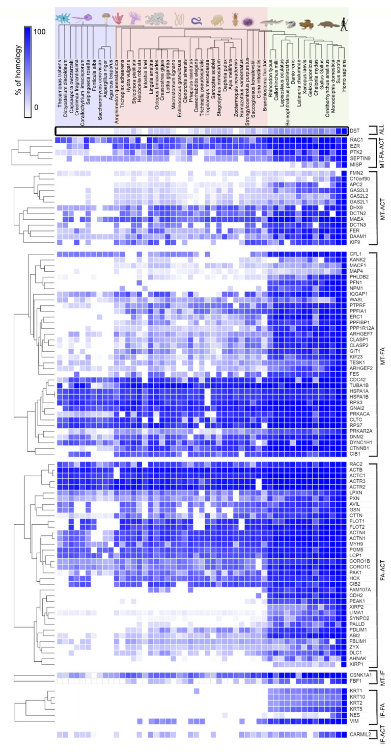
Adhesion between cells and the extracellular matrix (ECM) is one of the prerequisites for multicellularity, motility, and tissue specialization. Focal adhesions (FAs) are defined as protein complexes that mediate signals from the ECM to major components of the cytoskeleton (microtubules, actin, and intermediate filaments), and their mutual communication determines a variety of cellular processes. In this study, human cytoskeletal crosstalk proteins were identified by comparing datasets with experimentally determined cytoskeletal proteins. The spectraplakin dystonin was the only protein found in all datasets. Other proteins (FAK, RAC1, septin 9, MISP, and ezrin) were detected at the intersections of FAs, microtubules, and actin cytoskeleton. Homology searches for human crosstalk proteins as queries were performed against a predefined dataset of proteomes. This analysis highlighted the importance of FA communication with the actin and microtubule cytoskeleton, as these crosstalk proteins exhibit the highest degree of evolutionary conservation. Finally, phylogenetic analyses elucidated the early evolutionary history of spectraplakins and cortical microtubule stabilization complexes (CMSCs) as model representatives of the human cytoskeletal crosstalk. While spectraplakins probably arose at the onset of opisthokont evolution, the crosstalk between FAs and microtubules is associated with the emergence of metazoans. The multiprotein complexes contributing to cytoskeletal crosstalk in animals gradually gained in complexity from the onset of metazoan evolution.

Keywords: actin; cortical microtubule stabilization complex; cytoskeletal crosstalk; dystonin; evolution; focal adhesion; intermediate filaments; microtubule; spectraplakin.


Conflict of interest statement

The authors declare that the research was conducted in the absence of any commercial or financial relationships that could be construed as a potential conflict of interest.

Figures

Figure 1
Figure 1
Putative cytoskeletal crosstalk proteins in humans. Venn diagram depicting 100 common proteins when using different datasets related to microtubules (MT), focal adhesions (FA), intermediate filaments (IF), and actin filaments. Dystonin (purple) was found in all datasets, whereas crosstalk proteins found in the MT, FA, and actin datasets are shown in a dark blue rectangle. Proteins at the crossing points between MT and FA (pink), MT and actin (green), FA and actin (beige), IF and MT (light blue), IF and actin (red), IF and FA (yellow) were generated as described in the Section 4.
Figure 2
Figure 2
Schematic representation of vertical and horizontal crosstalk. (A) After binding to ECM proteins, the signal is transduced to focal adhesions via integrins. Proteins of the cortical microtubule stabilization complex (liprins, ERC1, PHLDB2, CLASPs, AMER2, and APC) play an important role in force transmission to microtubules, while direct binding of KANK to talin-1 and KIF21A contributes to microtubule recruitment to the outer rim of focal adhesions. (B) Force transmission to actin filaments is facilitated by the binding of talin-1 to cytoplasmic integrin tails that recruit mediators such as vinculin and actinin alpha and help to transduce the signal to actin filaments. (C) ECM-integrin-mediated force transmission to intermediate filaments is facilitated by binding of the β4-subunit and plectin, whereas depletion of vimentin blocks β4-enhanced invasion. In addition, binding of plectin to the β4 subunit is responsible for recruitment to focal adhesions and cell spreading. (D) Simplified scheme of vertical and horizontal crosstalk.
Figure 3
Figure 3
Evolutionary conservation of cytoskeletal crosstalk proteins. Heatmap representing percentage of homology for crosstalk proteins found at the crossing points of the microtubules (MT), focal adhesions (FAs), intermediate filaments (IFs), and actin (ACT) in human cytoskeleton datasets. The proteins found at the crossing points were classified according to their occurrence in the respective datasets, and hierarchical clustering (Euclidean distance algorithm and average linkage method) was performed for each group separately using the Morpheus online tool.
Figure 4
Figure 4
Analysis of spectraplakin proteins and domains in animals, and its closest relatives. (A) Phylogenetic relationship of different spectrin, spectraplakin, and GAS2 proteins. The phylogenetic tree was reconstructed based on concatenated CH, GAS2, SR1 and SR4 (or the last SR in spectrins, spectraplakins and SR-GAS fungal proteins) protein domains using a maximum likelihood method. The numbers indicate SH-aLRT and UFBoot values (only >80 values are shown). Tree is midpoint rooted. Schematic domain representation of corresponding proteins is designated on the right: CH —calponin homology domain, PR—plectin repeat, SR—spectrin repeat, GAS2—growth arrest specific 2 (Gas2)-related domain, SH3—Src-homology 3 domain. (B) GAS domain conservation. GAS domains from spectraplakins and GAS proteins belonging to phylogenetically distant organisms were extracted using InterProScan and aligned. β plates 3, 4, and 5 indicate MT interacting site. (C) SH3 (Src-homology 3) motif conservation in spectrin and spectraplakin proteins. Alignment of SH3 motifs from spectraplakin and spectrin proteins were identified by InterProScan. Putative interacting sites are designated with red arrows. Red boxes indicate change from spectrin aromatic residues to smaller side-chain amino acids. Overall conservation is indicated below alignment, (B,C) 0→* increasing amino acid (aa) residues conservation, * = aa residues in all sequences are conserved. (D) Plectin repeat domain (PR) was identified by InterProScan in human spectraplakins/plakins and different Protostomian spectraplakins. Identification of conserved repeated motifs was obtained by MEME suite. (E) Proposed evolutionary scenario of spectraplakins and their domains and other related proteins.
Figure 5
Figure 5
Analysis of evolutionary origins of crosstalk proteins found at intersection of 3 cytoskeletal protein datasets. (A) Evolutional emergence of crosstalk proteins found in 3 datasets starting with RAC1 (blue), followed by septin 9 (red), FAK (purple), ezrin (yellow) and MISP (green) as the newest cytoskeletal crosstalk player detected in this analysis. Although ezrin is evolutionary young protein, ERM domain is present before Metazoa. (B) Identification of conserved repeated motifs was obtained by MEME suite; motif consensuses are designated in upper right box. Phylogenetic and conserved domains analysis of MISP and its homologs in different Chordata species (lower box). ML tree of MISP homologs, aLRT values are used to infer branch support, values on the main branches are indicated. Phallusia mammilata and Ciona intestinalis (Chordata, Tunicata) homologs are placed as outgroup. Sequences IDs are listed in Supplementary Table S1 (Sheet 7).
Figure 6
Figure 6
Analysis of the evolutionary origins of CMSC complexes as microtubule-focal adhesion linkers. (A) Presence of cortical microtubule stabilisation complex (CMSC) component homologs in different Amorphea groups. The search for homologs was performed as described in M&M, hits obtained after initial BLAST search to human queries were reciprocally BLAST to verify the best hits. A protein was marked as a partial homolog if the coverage was <40% compared to the human protein, while in the case of KIF21A and KANK, conservation of known interaction motifs with KANK and talin was used, respectively. (B) Conservation of KN motif in KANK (talin-binding), residues involved in specific interactions with talin are marked with red line. (C) Conservation of KBD (KANK-binding domain) in KIF21A, residues involved in specific interactions with KANK are marked with red/blue arrows. (B,C) 0→* increasing amino acid (aa) residues conservation, * = aa residues in all sequences are conserved. (D) Increase in complexity of CMSC components from unicellular opisthokonts to Chordata. Although CMSC originate in the premetazoa, they gradually acquire new components during animal evolution.
Figure 7
Figure 7
Suggested pathway involving five proteins found at the intersections of FA, MT and actin filament datasets. The hypothetical mechanism involves RAC1, septin 9, FAK, ezrin and MISP depicted together with common interacting partners.

Similar articles

Cited by

References

    1. Jaalouk D.E., Lammerding J. Mechanotransduction gone awry. Nat. Rev. Mol. Cell Biol. 2009;10:63–73. doi: 10.1038/nrm2597. - DOI - PMC - PubMed
    1. Harjunpää H., Llort Asens M., Guenther C., Fagerholm S.C. Cell Adhesion Molecules and Their Roles and Regulation in the Immune and Tumor Microenvironment. Front. Immunol. 2019;10:1078. doi: 10.3389/fimmu.2019.01078. - DOI - PMC - PubMed
    1. Samanta D., Almo S.C. Nectin family of cell-adhesion molecules: Structural and molecular aspects of function and specificity. Cell. Mol. Life Sci. 2015;72:645–658. doi: 10.1007/s00018-014-1763-4. - DOI - PMC - PubMed
    1. Hynes R.O. Integrins: A family of cell surface receptors. Cell. 1987;48:549–554. doi: 10.1016/0092-8674(87)90233-9. - DOI - PubMed
    1. Hynes R.O. Integrins: Bidirectional, Allosteric Signaling Machines. Cell. 2002;110:673–687. doi: 10.1016/S0092-8674(02)00971-6. - DOI - PubMed