Review
. 2022 May 21;23(10):5769.
doi: 10.3390/ijms23105769.

Dynamic Involvement of Telocytes in Modulating Multiple Signaling Pathways in Cardiac Cytoarchitecture

Affiliations
Review

Dynamic Involvement of Telocytes in Modulating Multiple Signaling Pathways in Cardiac Cytoarchitecture

Ioana Cucu et al. Int J Mol Sci. .

Abstract

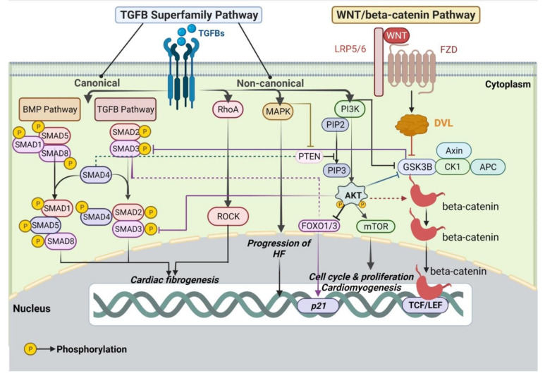
Cardiac interstitium is a complex and dynamic environment, vital for normal cardiac structure and function. Telocytes are active cellular players in regulating main events that feature myocardial homeostasis and orchestrating its involvement in heart pathology. Despite the great amount of data suggesting (microscopically, proteomically, genetically, etc.) the implications of telocytes in the different physiological and reparatory/regenerative processes of the heart, understanding their involvement in realizing the heart's mature cytoarchitecture is still at its dawn. Our scrutiny of the recent literature gave clearer insights into the implications of telocytes in the WNT signaling pathway, but also TGFB and PI3K/AKT pathways that, inter alia, conduct cardiomyocytes differentiation, maturation and final integration into heart adult architecture. These data also strengthen evidence for telocytes as promising candidates for cellular therapies in various heart pathologies.

Keywords: PI3K/AKT; TGFB; WNT; heart failure; telocytes.


Conflict of interest statement

The authors declare no conflict of interest.

Figures

Figure 1
Figure 1
An illustrative representation of the interlinking networks between WNT and TGFB signaling pathways. In the sketch are delineated the intricate protein–protein interaction latticeworks within cardiomyocytes and telocytes, showing a complementary synergism to induce cardiomyogenesis, cardiac regeneration, and pathogenesis of heart diseases. The figure is widely scrutinized in the main text. The black arrows designate main signaling pathways; the dashed arrows indicate activation; the continuous blunt-ended arrows pinpoint inhibition. APC: Adenomatous polyposis coli; BetaTrCP: Beta transducin repeats-containing protein; BMP: Bone morphogenetic protein; CAMKII: Calcium calmodulin-dependent protein kinase II; CK1: Casein kinase 1; DAAM: Disheveled-associated activator of morphogenesis; DAG: Diacylglycerol; DKK: Dickkopf; DVL: Dishevelled; FZD: Frizzled; GSK3B: Glycogen synthase kinase 3 beta; IP3: Inositol trisphosphate; JNK: c-Jun N terminal kinase; LGR5: Leucine-rich repeat-containing G-protein coupled receptor; LRP5/6: Lipoprotein receptor-related protein 5/6; NFAT: Nuclear factor of activated T-cells; PKC: Protein kinase C; PLC: Phospholipase C; RNF43: Ring finger protein 43; ROCK: Rho-associated protein kinase; RSPO: R-spondin; SMAD: Small mothers against decapentaplegic; TAK1/NLK: Transforming growth factor beta-activated kinase 1/Nemo like kinase; TCF/LEF: T cell factor/Lymphoid enhancer factor; TGFB: Transforming growth factor beta; sFRP: Secreted Frizzled-related proteins; ZNRF3: Zinc and ring finger protein 3. Created with https://biorender.com/, accessed on 30 April 2022.
Figure 2
Figure 2
Illustration of the interconnecting networks between WNT, TGFB, PI3K/AKT/mTOR, and MAPK pathways. The figure is broadly discussed in the main text. The black arrows designate main signaling pathways; the dashed arrows indicate activation; the continuous blunt-ended arrows pinpoint inhibition. AKT: v-akt murine thymoma viral oncogene homolog/Protein kinase B; APC: Adenomatous polyposis coli; BMP: Bone morphogenetic protein; CK1: Casein kinase 1; FOXO1/3: Forkhead box proteins O1/3; FZD: Frizzled; GSK3B: Glycogen synthase kinase 3 beta; HF: heart failure; LRP5/6: Lipoprotein receptor-related protein 5/6; MAPK: Mitogen-activated protein kinase; mTOR: mammalian target of rapamycin; PI3K: phosphatidylinositol 3-kinase; PIP2: Phosphatidylinositol-4,5-bisphosphate; PIP3: Phosphatidylinositol-3,4,5-triphosphate; PTEN: Phosphatase and tensin homolog deleted on chromosome 10; ROCK: Rho-associated protein kinase; SMAD: Small mothers against decapentaplegic; TCF/LEF: T cell factor/Lymphoid enhancer factor; TGFB: Transforming growth factor beta. Created with https://biorender.com/, accessed on 30 April 2022.

References

    1. Popescu L.M., Gherghiceanu M., Hinescu M.E., Cretoiu D., Ceafalan L., Regalia T., Popescu A.C., Ardeleanu C., Mandache E. Insights into the interstitium of ventricular myocardium: Interstitial Cajal-like cells (ICLC) J. Cell. Mol. Med. 2006;10:429–458. doi: 10.1111/j.1582-4934.2006.tb00410.x. - DOI - PMC - PubMed
    1. Etoh T., Joffs C., Deschamps A.M., Davis J., Dowdy K., Hendrick J., Baicu S., Mukherjee R., Manhaini M., Spinale F.G. Myocardial and interstitial matrix metalloproteinase activity after acute myocardial infarction in pigs. Am. J. Physiol. Heart Circ. Physiol. 2001;281:H987–H994. doi: 10.1152/ajpheart.2001.281.3.H987. - DOI - PubMed
    1. Iles L., Pfluger H., Phrommintikul A., Cherayath J., Aksit P., Gupta S.N., Kaye D.M., Taylor A.J. Evaluation of diffuse myocardial fibrosis in heart failure with cardiac magnetic resonance contrast-enhanced T1 mapping. J. Am. Coll. Cardiol. 2008;52:1574–1580. doi: 10.1016/j.jacc.2008.06.049. - DOI - PubMed
    1. Popescu L.M., Faussone-Pellegrini M.-S. TELOCYTES—a case of serendipity: The winding way from Interstitial Cells of Cajal (ICC), via Interstitial Cajal-Like Cells (ICLC) to TELOCYTES. J. Cell. Mol. Med. 2010;14:729–740. doi: 10.1111/j.1582-4934.2010.01059.x. - DOI - PMC - PubMed
    1. Cretoiu S.M., Popescu L.M. Telocytes revisited. Biomol. Concepts. 2014;5:353–369. doi: 10.1515/bmc-2014-0029. - DOI - PubMed

LinkOut - more resources