. 2022 May 21;23(10):5788.
doi: 10.3390/ijms23105788.

Towards 3D Bioprinted Spinal Cord Organoids

Affiliations

Towards 3D Bioprinted Spinal Cord Organoids

Yilin Han et al. Int J Mol Sci. .

Abstract

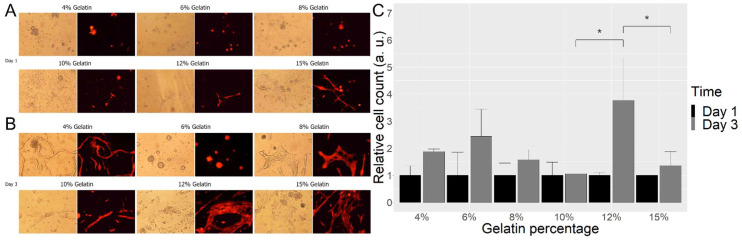
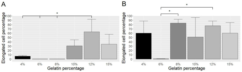
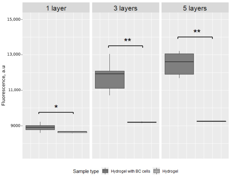
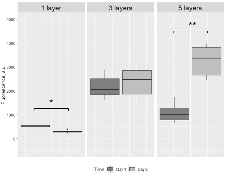
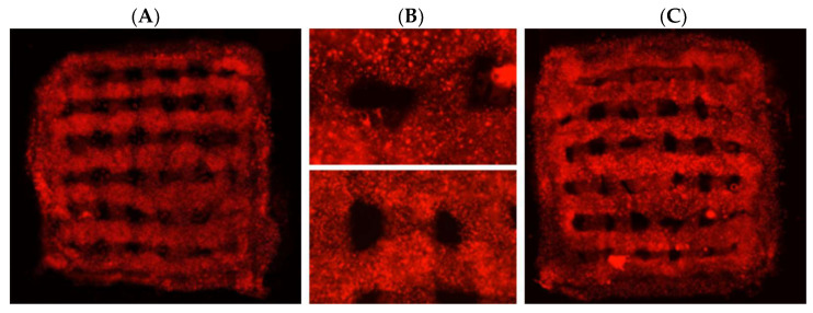
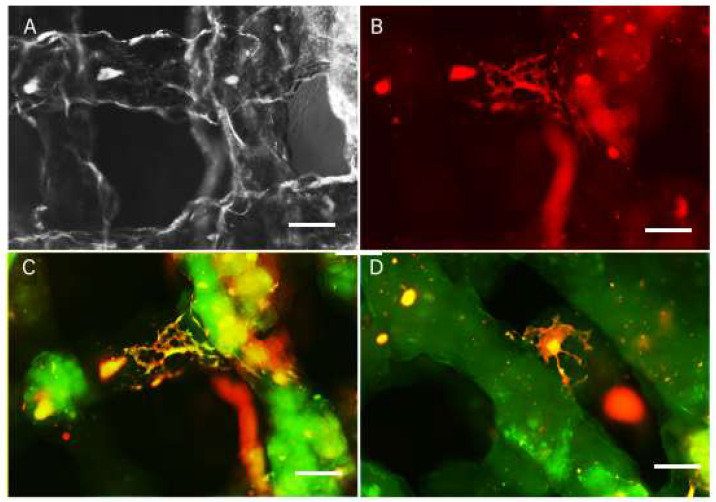
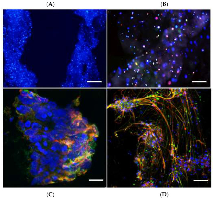
Three-dimensional (3D) cultures, so-called organoids, have emerged as an attractive tool for disease modeling and therapeutic innovations. Here, we aim to determine if boundary cap neural crest stem cells (BC) can survive and differentiate in gelatin-based 3D bioprinted bioink scaffolds in order to establish an enabling technology for the fabrication of spinal cord organoids on a chip. BC previously demonstrated the ability to support survival and differentiation of co-implanted or co-cultured cells and supported motor neuron survival in excitotoxically challenged spinal cord slice cultures. We tested different combinations of bioink and cross-linked material, analyzed the survival of BC on the surface and inside the scaffolds, and then tested if human iPSC-derived neural cells (motor neuron precursors and astrocytes) can be printed with the same protocol, which was developed for BC. We showed that this protocol is applicable for human cells. Neural differentiation was more prominent in the peripheral compared to central parts of the printed construct, presumably because of easier access to differentiation-promoting factors in the medium. These findings show that the gelatin-based and enzymatically cross-linked hydrogel is a suitable bioink for building a multicellular, bioprinted spinal cord organoid, but that further measures are still required to achieve uniform neural differentiation.

Keywords: bioprinting; cell differentiation; cell survival; hydrogel; neural stem cell.


Conflict of interest statement

The authors declare no conflict of interest.

Figures

Figure 1
Figure 1
Images of BC on scaffolds from day 1 (A) and day 3 (B) with different concentrations of gelatin. Relative cell number on different scaffolds at day 1 and day 3 (C). Cell numbers on different concentrations of gelatin were assessed on day 1 and day 3 after attachment. For each concentration of gelatin, three random images of cultures were taken, and the number of cells estimated by ImageJ, via virtual measurement. Data and means are from three independent experiments. * p < 0.01. Scale bar A,B = 100 microns. Additional image with the scale bar.
Figure 2
Figure 2
Differentiation of BC on gelatin of different concentrations after one day (A) and three days (B) of culture. The proportion of elongated cells was calculated by counting the cell numbers of non-elongated and elongated cells in three random images. * p ≤ 0.05.
Figure 3
Figure 3
Box plot of the fluorescence intensity after 72 h of incubation of cell-loaded scaffolds (hydrogel with BC cells) and control scaffolds (hydrogel only) printed in one, three or five layers. * p < 0.01, ** p < 0.0001.
Figure 4
Figure 4
Box plot of the fluorescence intensity after 24 and 72 h of cell-loaded scaffolds, printed in one, three or five layers. * p < 0.01, ** p < 0.0001, ● - outlier. There is a significant decrease in fluorescence intensity from 24 to 72 h in one-layered structures, indicating loss of viable cells. The significant increase in fluorescence intensity from 24 to 72 h in five-layered structures presumably reflects a slower medium penetration through this structure compared to three-layered structures.
Figure 5
Figure 5
3D-printed bioscaffolds with red-fluorescent BC cells. (A) and (B) upper panel—day 1 after printing. (B) Lower panel and (C)—5 weeks after printing. The relocation of cells towards the edge of the printed structure in 5 weeks bioscaffolds is shown.
Figure 6
Figure 6
Live images of scaffolds (A,B) and fixed scaffolds 3 weeks after printing. GFAP staining (green) reveals glial cells differentiated in the scaffold (C) and bTUB staining (D) reveals neurons differentiated in the scaffold, with the cell bodies located inside the scaffold and on the surface of the printed structure. Scale bar: AD = 10 microne.
Figure 7
Figure 7
Immunofluorescence labeling of iPSC-derived neural cells after 1 week and 3 weeks of culture inside the scaffold (A,B) and on the surface of scaffold (C,D). Sections are labelled with antibodies to bTUB (green) and GFAP (red), and stained with Hoechst (blue). (A) Cells are distributed throughout the scaffold. (B) Inside the scaffold, the cells express neural markers, but do not extend processes. (C) In the peripheral part of the scaffold, after 1 week in culture, there are differentiated neurons and astrocytes on the surface of the scaffold. (D) After three weeks in culture, there is extensive neurite formation from neurons, intermingled with astrocytic processes on the surface of the scaffolds. Scale bar: A = 20 microns; B = 10 microns; C = 5 microns; D = 10 microns.

References

    1. Bose R., Banerjee S., Dunbar G.L. Modeling Neurological Disorders in 3D Organoids Using Human-Derived Pluripotent Stem Cells. Front. Cell Dev. Biol. 2021;9:640212. doi: 10.3389/fcell.2021.640212. - DOI - PMC - PubMed
    1. Porciúncula L.O., Goto-Silva L., Ledur P.F., Rehen S.K. The Age of Brain Organoids: Tailoring Cell Identity and Functionality for Normal Brain Development and Disease Modeling. Front. Neurosci. 2021;15:674563. doi: 10.3389/fnins.2021.674563. - DOI - PMC - PubMed
    1. Martins J.-M.F., Fischer C., Urzi A., Vidal R., Kunz S., Ruffault P.-L., Kabuss L., Hube I., Gazzerro E., Birchmeier C., et al. Self-Organizing 3D Human Trunk Neuromuscular Organoids. Cell Stem Cell. 2020;26:172–186.e6. doi: 10.1016/j.stem.2019.12.007. - DOI - PubMed
    1. Andersen J., Revah O., Miura Y., Thom N., Amin N.D., Kelley K.W., Singh M., Chen X., Thete M.V., Walczak E.M., et al. Generation of Functional Human 3D Cortico-Motor Assembloids. Cell. 2020;183:1913–1929.e26. doi: 10.1016/j.cell.2020.11.017. - DOI - PMC - PubMed
    1. Hor J.-H., Ng S.-Y. Methods in Cell Biology. Volume 159. Academic Press; Cambridge, MA, USA: 2020. Generating ventral spinal organoids from human induced pluripotent stem cells; pp. 257–277. - DOI - PubMed

LinkOut - more resources