. 2022 May 12;11(10):2740.
doi: 10.3390/jcm11102740.

Adiponectin Is a Component of the Inflammatory Cascade in Rheumatoid Arthritis

Affiliations

Adiponectin Is a Component of the Inflammatory Cascade in Rheumatoid Arthritis

Małgorzata Łączna et al. J Clin Med. .

Abstract

Adiponectin is a secretory protein of adipocytes that plays an important role in pathological processes by participation in modulating the immune and inflammatory responses. The pro-inflammatory effect of adiponectin is observed in rheumatoid arthritis (RA). In this study, we examined adiponectin plasma levels and the expression of adiponectin in bone marrow tissue samples, synovium samples, and infrapatellar fat pad samples from patients with osteoarthritis (OA) and RA. Additionally we examined the expression of adiponectin receptors AdipoR1 and AdipoR2 in synovium samples and infrapatellar fat pad samples from patients with OA and RA. We also assessed the correlations between adiponectin plasma concentrations, adiponectin expression in bone marrow, synovium, infrapatellar fat pad, and plasma levels of selected cytokines. We found increased expression of adiponectin in synovium samples and infrapatellar fat pad samples from patients with RA as compared to patients with OA. There were no statistically significant differences of adiponectin plasma levels and adiponectin expression in bone marrow tissue samples between OA and RA patients. There were no differences in the expression of AdipoR1 and AdipoR2 at the mRNA level in synovial tissue and the infrapatellar fat pad between RA and OA patients. However, in immunohistochemical analysis in samples of the synovial membrane from RA patients, we observed very strong expression of adiponectin in intima cells, macrophages, and subintimal fibroblasts, such as synoviocytes, vs. strong expression in OA samples. Very strong expression of adiponectin was also noted in adipocytes of Hoffa's fat pad of RA patients. Expression of AdipoR1 was stronger in RA tissue samples, while AdipoR2 expression was very similar in both RA and OA samples. Our results showed increased adiponectin expression in the synovial membrane and Hoffa's pad in RA patients compared to that of OA patients. However, there were no differences in plasma adiponectin concentrations and its expression in bone marrow. The results suggest that adiponectin is a component of the inflammatory cascade that is present in RA. Pro-inflammatory factors enhance the expression of adiponectin, especially in joint tissues-the synovial membrane and Hoffa's fat pad. In turn, adiponectin also increases the expression of further pro-inflammatory mediators.

Keywords: adiponectin; bone marrow; infrapatellar fat pad; rheumatoid arthritis; synoviocytes.


Conflict of interest statement

The authors declare that the research was conducted in the absence of any commercial or financial relationships that could be construed as a potential conflict of interest.

Figures

Figure 1
Figure 1
Plasma adiponectin concentrations at the protein level (mean ± SD, µg/mL) in OA—osteoarthritis and RA—rheumatoid arthritis patients and in healthy subjects (HC—healthy controls) (Kruskal–Wallis test).
Figure 2
Figure 2
mRNA expression levels of adiponectin (ADIPOQ) in OA—osteoarthritis and RA—rheumatoid arthritis patients (mean ± SD): Panel (A) infrapatellar fat pad tissue, Panel (B) synovium, Panel (C) bone marrow. * p < 0.05 indicates a significant increase in gene expression in RA patients as compared to OA patients (Mann–Whitney test).
Figure 3
Figure 3
mRNA expression levels of adiponectin receptor 1 (ADIPOR1) and adiponectin receptor 2 (ADIPOR2) in OA—osteoarthritis and RA—rheumatoid arthritis patients (mean ± SD): Panel (A,C) infrapatellar fat pad tissue, Panel (B,D) synovium. There were no significant differences in ADIPOR1 and ADIPOR2 gene expression in RA patients as compared to OA patients (Mann–Whitney test).
Figure 4
Figure 4
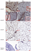
Adiponectin expression in joint’s tissues. Panels (AC) synovial membranes; Panels (DF) Hoffa’s fat pad. OA—osteoarthritis; RA—rheumatoid arthritis; V—vessel; Panels (C,F) show negative control staining. Objective magnification ×20, scale bar 50 μm; only representative images are presented. Insets show large magnifications of cytoplasmic staining in synovial membrane cells (Panel (B)) and endothelial cells (Panel (A)).
Figure 5
Figure 5
AdipoR1 and AdipoR2 expressions in joint tissues: Panels (AD) synovial membranes; Panels (EH) Hoffa’s fat pads. OA—osteoarthritis; RA—rheumatoid arthritis; Panels (I,J) show negative control staining. Insert shows gathered inflammatory cells positively stained for AdipoR2. Objective magnification ×20, scale bar 50 μm; only representative images are presented. Inserts show large magnification of cytoplasmic staining in synovial membrane cells—Panels (B,C), and immune cells staining—Panel (D).
Figure 6
Figure 6
Effect of stimulation of human fibroblast-like synoviocytes (HFLSs) with LPS, TNF-α, and LPS + TNF-α on adiponectin mRNA expression. Means from 3 experiments ± 95% confidence intervals are presented. * p < 0.05 (Tukey post hoc test in comparison to non-stimulated control). TNF: Tumor necrosis factor. LPS: Lipopolysaccharide.
Figure 7
Figure 7
The effect of adiponectin (ADIPO) stimulation of human fibroblast-like synoviocytes (HFLSs) on IL-6 mRNA expression. Red arrow indicates negative control (without adiponectin and without LPS). Black arrow indicates control with LPS alone (without adiponectin). Means from 3 experiments ± 95% confidence intervals are presented. * p < 0.05 (Tukey post hoc test in comparison to cells not stimulated with adiponectin).

References

    1. Obata Y., Yamada Y., Takahi Y., Baden M.Y., Saisho K., Tamba S., Yamamoto K., Umeda M., Furubayashi A., Matsuzawa Y. Relationship between serum adiponectin levels and age in healthy subjects and patients with type 2 diabetes. Clin. Endocrinol. 2013;79:204–210. doi: 10.1111/cen.12041. - DOI - PubMed
    1. Achari A.E., Jain S.K. Adiponectin, a Therapeutic Target for Obesity, Diabetes, and Endothelial Dysfunction. Int. J. Mol. Sci. 2017;18:61321. doi: 10.3390/ijms18061321. - DOI - PMC - PubMed
    1. Guerre-Millo M. Adipose tissue hormones. J. Endocrinol. Investig. 2002;25:855–861. doi: 10.1007/BF03344048. - DOI - PubMed
    1. Cook W.S., Yeldandi A.V., Rao M.S., Hashimoto T., Reddy J.K. Less extrahepatic induction of fatty acid beta-oxidation enzymes by PPAR alpha. Biochem. Biophys. Res. Commun. 2000;278:250–257. doi: 10.1006/bbrc.2000.3739. - DOI - PubMed
    1. Choi H.M., Doss H.M., Kim K.S. Multifaceted Physiological Roles of Adiponectin in Inflammation and Diseases. Int. J. Mol. Sci. 2020;21:41219. doi: 10.3390/ijms21041219. - DOI - PMC - PubMed

LinkOut - more resources