. 2022 May 17;11(10):2832.
doi: 10.3390/jcm11102832.

Flow Cytometry Analysis of Blood Large Extracellular Vesicles in Patients with Multiple Sclerosis Experiencing Relapse of the Disease

Affiliations

Flow Cytometry Analysis of Blood Large Extracellular Vesicles in Patients with Multiple Sclerosis Experiencing Relapse of the Disease

Jakub Soukup et al. J Clin Med. .

Abstract

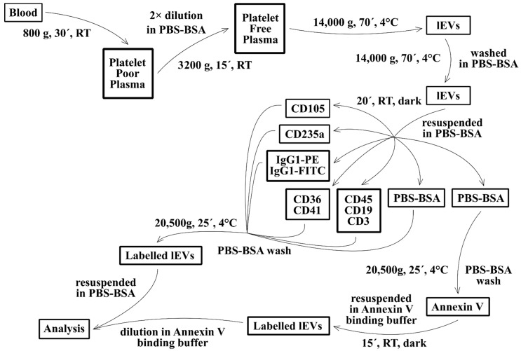
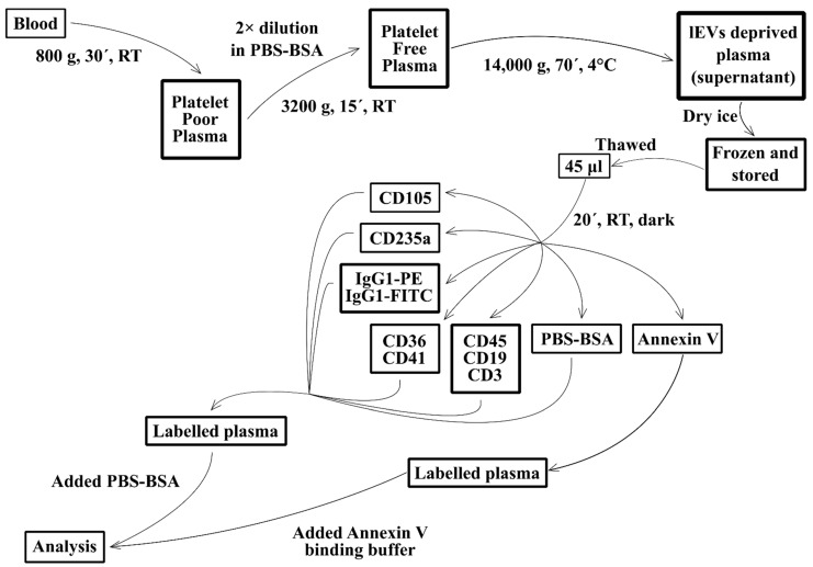
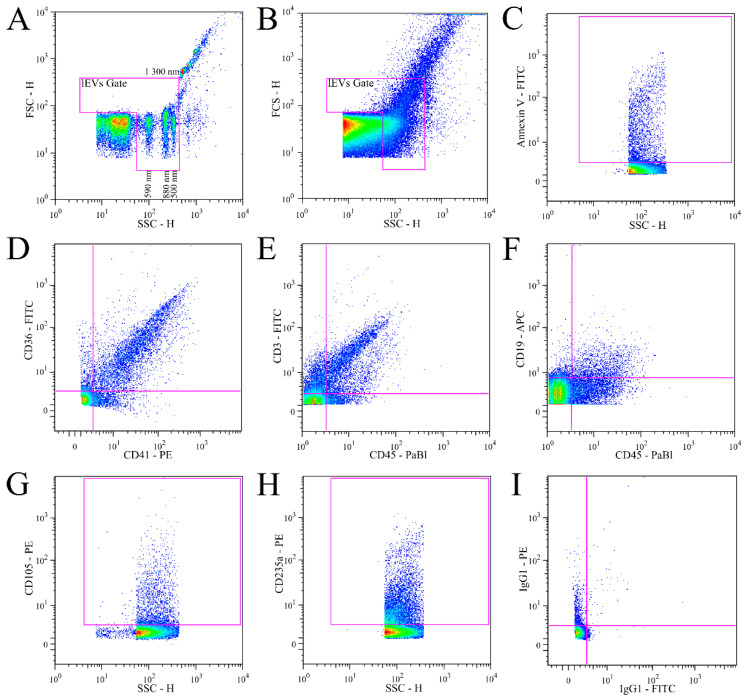
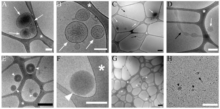
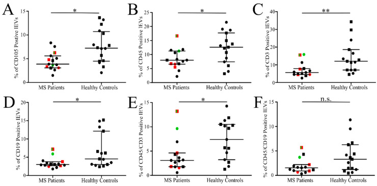
The number of people living with multiple sclerosis (MS) in developed countries is increasing. The management of patients is hindered by the absence of reliable laboratory tests accurately reflecting the disease activity. Extracellular vesicles (EVs) of different cell origin were reportedly elevated in MS patients. We assessed the diagnostic potential, with flow cytometry analysis, of fresh large EVs (lEVs), which scattered more light than the 590 nm silica beads and were isolated from the blood plasma of relapsing remitting MS patients. Venous blood was collected from 15 patients and 16 healthy controls (HC). The lEVs were isolated from fresh platelet-free plasma by centrifugation, labelled with antibodies and the presence of platelet (CD41+, CD36+), endothelial (CD105+), erythrocyte (CD235a+), leukocyte (CD45+, CD19+, CD3+) and phosphatidylserine (Annexin V+) positive lEVs was analyzed using standard flow cytometry. Cryo-electron microscopy was used to verify the presence of EVs in the analyzed plasma fractions. MS patients experiencing acute relapse had slightly reduced relative levels (% of positive lEVs) of CD105+, CD45+, CD3+, CD45+CD3+ or CD19+ labelled lEVs in comparison to healthy controls. An analysis of other markers or a comparison of absolute lEV counts (count of lEVs/µL) did not yield any significant differences. Our data do not support the hypothesis that the exacerbation of the disease in RRMS patients leads to an increased numbers of circulating plasma lEVs which can be monitored by standard flow cytometry.

Keywords: cryo-electron microscopy; extracellular vesicles; flow cytometry; multiple sclerosis; plasma.


Conflict of interest statement

The authors declare no conflict of interest.

Figures

Figure 1
Figure 1
Scheme of isolation and labelling of fresh large extracellular vesicles (lEVs) from blood. Each time one patient and one healthy control blood sample were processed simultaneously. RT—room temperature; PBS-BSA—phosphate buffered saline pH 7.4 with 0.1% bovine serum albumin.
Figure 2
Figure 2
Scheme of preparation and labelling of plasma deprived of lEVs by centrifugation. Each time one patient and one healthy control plasma sample were processed simultaneously. RT—room temperature; PBS-BSA—phosphate buffered saline pH 7.4 with 0.1% bovine serum albumin.
Figure 3
Figure 3
Definition of lEVs gate and representative results of lEVs labelling. (A). ApogeeMix beads were used to set lEVs gate to size of the beads. The mix consisted of 180 nm, 240 nm, 300 nm (below sensitivity), 590 nm, 880 nm and 1,300 nm silica beads with refractive index 1.43 and 2 latex beads (110 nm and 500 nm) with green fluorescence and refractive index 1.59. (B). An illustrative scattergram of isolated plasma vesicles with lEVs gate. Only events in the lEVs gate were included in the analysis. (CI). Illustrative fluorescence dot plots showing representative results of lEVs labelling with: Annexin V FITC (C), CD36 FITC + CD41 PE (D), CD45 PaBl + CD3 FITC (E), CD45 PaBl + CD19 APC (F), CD105 PE (G), CD235a PE (H) and isotypic control IgG1 FITC + IgG1 PE (I).
Figure 4
Figure 4
Cryo-TEM of isolated lEVs (upper row) and supernatant plasma deprived of lEVs (lower row). (A,B) A detailed picture of lEVs (white arrow). (C). Overview of lEVs sample. lEVs (white arrow), elongated vesicle (black arrow) (D). Elongated vesicles (black arrow) which occasionally present in isolated lEVs (white arrow). (E). Electron-dense vesicles commonly present in plasma supernatant after lEVs isolation (white arrowhead). (F). A detailed picture of the electron-dense vesicle (white arrowhead). (G). Plasma supernatant showing the electron-dense vesicles (white arrowheads) and impurities such as protein aggregates. (H). Small (up to 50 nm) vesicles (black arrowhead) and proteins are visible in the picture. White scale bar 200 nm, black scale bar 500 nm. Asterisk marks lacey carbon support film on the grid.
Figure 5
Figure 5
Relative number of labelled isolated lEVs in MS patients and HC. Percentage of endothelial CD105+ (A), leukocyte CD45+ (B), T-lymphocyte CD3+ (C) and CD45+CD3+ (E), B-lymphocyte CD19+ (D) and CD45+CD19+ (F) lEVs out of all events collected in lEVs gate. The line represents median value with interquartile range. Women (circles), men (squares), patients without treatment (green), patients receiving intravenous corticoids up to 14 days before blood collection (red). * p < 0.05, ** p < 0.005, n.s.—not significant; MS patients (n = 15) and HC (n = 15 or n = 16).

References

    1. Thompson A.J., Baranzini S.E., Geurts J., Hemmer B., Ciccarelli O. Multiple sclerosis. Lancet. 2018;391:1622–1636. doi: 10.1016/S0140-6736(18)30481-1. - DOI - PubMed
    1. Saposnik G., Sempere A.P., Raptis R., Prefasi D., Selchen D., Maurino J. Decision making under uncertainty, therapeutic inertia, and physicians’ risk preferences in the management of multiple sclerosis (DIScUTIR MS) BMC Neurol. 2016;16:9. doi: 10.1186/s12883-016-0577-4. - DOI - PMC - PubMed
    1. Trapp B.D., Nave K.A. Multiple sclerosis: An immune or neurodegenerative disorder? Annu. Rev. Neurosci. 2008;31:247–269. doi: 10.1146/annurev.neuro.30.051606.094313. - DOI - PubMed
    1. Nylander A., Hafler D.A. Multiple sclerosis. J. Clin. Investig. 2012;122:1180–1188. doi: 10.1172/JCI58649. - DOI - PMC - PubMed
    1. Raposo G., Stoorvogel W. Extracellular vesicles: Exosomes, microvesicles, and friends. J. Cell Biol. 2013;200:373–383. doi: 10.1083/jcb.201211138. - DOI - PMC - PubMed