Review
. 2022 May 19;11(10):2866.
doi: 10.3390/jcm11102866.

The Applications of Artificial Intelligence in Cardiovascular Magnetic Resonance-A Comprehensive Review

Affiliations
Review

The Applications of Artificial Intelligence in Cardiovascular Magnetic Resonance-A Comprehensive Review

Adriana Argentiero et al. J Clin Med. .

Abstract

Cardiovascular disease remains an integral field on which new research in both the biomedical and technological fields is based, as it remains the leading cause of mortality and morbidity worldwide. However, despite the progress of cardiac imaging techniques, the heart remains a challenging organ to study. Artificial intelligence (AI) has emerged as one of the major innovations in the field of diagnostic imaging, with a dramatic impact on cardiovascular magnetic resonance imaging (CMR). AI will be increasingly present in the medical world, with strong potential for greater diagnostic efficiency and accuracy. Regarding the use of AI in image acquisition and reconstruction, the main role was to reduce the time of image acquisition and analysis, one of the biggest challenges concerning magnetic resonance; moreover, it has been seen to play a role in the automatic correction of artifacts. The use of these techniques in image segmentation has allowed automatic and accurate quantification of the volumes and masses of the left and right ventricles, with occasional need for manual correction. Furthermore, AI can be a useful tool to directly help the clinician in the diagnosis and derivation of prognostic information of cardiovascular diseases. This review addresses the applications and future prospects of AI in CMR imaging, from image acquisition and reconstruction to image segmentation, tissue characterization, diagnostic evaluation, and prognostication.

Keywords: artificial intelligence; cardiac magnetic resonance; deep learning; machine learning.


Conflict of interest statement

The authors declare no conflict of interest.

Figures

Figure 1
Figure 1
The most commonly used Machine Learning (ML) techniques in the field of cardiac imaging.
Figure 2
Figure 2
LGE sequences acquired using an artificial intelligence reconstruction deep learning algorithm. Forty-one-year old patient with previous myocardial infarction on anterior, anteroseptal, inferoseptal, inferior, and inferolateral segments (arrows, (AE), respectively). Image noise decreased progressively with increase in AIRDL reconstruction in both 2D-SSLGE ((A): 2D-SSLGE AIRDL 0%, (B): 2D-SSLGE AIRDL 25%, (C): 2D-SSLGE AIRDL 50%, (D): 2D-SSLGE AIRDL 75%, (E): 2D-SSLGE AIRDL 100%). 2D-SSLGE—2D single segmented inversion recovery gradient echo late gadolinium enhancement sequences; AIRDL—artificial intelligence reconstruction deep learning.
Figure 3
Figure 3
Short axis acquired using parallel imaging and compressed sensing methods. (A) string shows a functional cardiac plane acquired using parallel imaging with a 1.5T MR system, while (B) string shows an SA acquired using the CS method with a 3T MR system. SA—short axis; CS—compressed sensing.
Figure 4
Figure 4
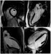
LGE —late gadolinium enhancement sequences acquired using the compressed sensing technique. Panel (A)—Left ventricle (LV) and right ventricle (RV) short axis view at the level pf the papillary muscles; (B)—LV three chamber view; (C)—LV two chamber view; (D)—LV and RV four chamber view.
Figure 5
Figure 5
Three-dimensional navigator whole-heart CMRA sequence. The 3D CMRA allows to acquire the whole heart in a gated free-breathing acquisition (A), with the possibility of subsequent MPR reconstructions (B,C). CMRA—coronary magnetic resonance angiography; MPR—Multiplanar reconstruction.
Figure 6
Figure 6
Artifacts reduction with artificial intelligence implementation. Eighty-two-year-old male patient with previous inferior and inferolateral myocardial infarction. Image (AC) show the reconstruction of 2D-MSLGE with NR 0% (A), NR 25% (B), and NR 50% (C), respectively. The increasing percentage in NR reconstruction yielded a progressive reduction in image noise in 2D-MSLGE starting from NR 0% (C) and moving through NR 25% (D) and NR 50% (E). A breath artifact characterizing the inferior and inferolateral midapical segments was reduced in the reconstruction in which the 100% artificial intelligence algorithm was applied. In fact, a reduction in quantum noise resulted in better contrast resolution. 2D-MSLGE—2D multisegment late gadolinium enhancement; NR—artificial intelligence reconstruction deep learning noise reduction.
Figure 7
Figure 7
Image segmentation platform. Functional and volume analysis to obtain ejection fraction, volumes, stroke volume, cardiac index, and left ventricle mass in end diastolic phase. Green: epicardial contour; red: endocardial contour; yellow: right ventricle. EDV—end diastolic volume; ESV—end systolic volume; SV—stroke volume; EF—ejection fraction; CO—cardiac output; CI—cardiac index.
Figure 8
Figure 8
Assessment of myocardial scar with semiautomatic tissue characterization. Semiautomatic tissue characterization algorithm allowing the identification of myocardial scars (represented by hyper-enhanced myocardium in panel (A) and yellow-colored myocardium in panel (B)) by positioning a region of interest within the territory of the remote/normal myocardium (dark myocardium). Green: epicardial contour; red: endocardial contour; yellow: myocardial scar; blue: normal myocardium.

Similar articles

Cited by

References

    1. Timmis A., Townsend N., Gale C.P., Torbica A., Lettino M., Petersen S.E., Mossialos E.A., Maggioni A.P., Kazakiewicz K., May H.T., et al. European Society of Cardiology Cardiovascular Disease Statistics 2019. Eur. Heart. J. 2020;41:12–85. doi: 10.1093/eurheartj/ehz859. - DOI - PubMed
    1. Pontone G., Andreini D., Bertella E., Baggiano A., Mushtaq S., Loguercio M., Segurini C., Conte E., Beltrama V., Annoni A., et al. Impact of an intra-cycle motion correction algorithm on overall evaluability and diagnostic accuracy of computed tomography coronary angiography. Eur. Radiol. 2016;26:147–156. doi: 10.1007/s00330-015-3793-1. - DOI - PubMed
    1. Guaricci A.I., Maffei E., Brunetti N.D., Montrone D., Di Biase L., Tedeschi C., Gentile G., Macarini L., Midiri M., Cademartiri F., et al. Heart rate control with oral ivabradine in computed tomography coronary angiography: A randomized comparison of 7.5 mg vs 5 mg regimen. Int. J. Cardiol. 2013;168:362–368. doi: 10.1016/j.ijcard.2012.09.041. - DOI - PubMed
    1. Pontone G., Weir-McCall J.R., Baggiano A., Del Torto A., Fusini L., Guglielmo M., Muscogiuri G., Guaricci A.I., Andreini D., Patel M., et al. Determinants of Rejection Rate for Coronary CT Angiography Fractional Flow Reserve Analysis. Radiology. 2019;292:597–605. doi: 10.1148/radiol.2019182673. - DOI - PubMed
    1. Pontone G., Andreini D., Guaricci A.I., Guglielmo M., Baggiano A., Muscogiuri G., Fusini L., Soldi M., Fazzari F., Berzovini C., et al. Quantitative vs. qualitative evaluation of static stress computed tomography perfusion to detect haemodynamically significant coronary artery disease. Eur. Heart. J. Cardiovasc. Imaging. 2018;19:1244–1252. doi: 10.1093/ehjci/jey111. - DOI - PubMed