

Artificial Intelligence-Enhanced Echocardiography for Systolic Function Assessment
- PMID: 35629019
- PMCID: PMC9143561
- DOI: 10.3390/jcm11102893
Artificial Intelligence-Enhanced Echocardiography for Systolic Function Assessment
Abstract
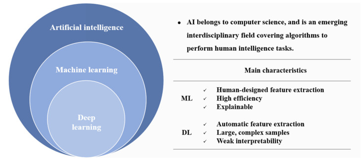
The accurate assessment of left ventricular systolic function is crucial in the diagnosis and treatment of cardiovascular diseases. Left ventricular ejection fraction (LVEF) and global longitudinal strain (GLS) are the most critical indexes of cardiac systolic function. Echocardiography has become the mainstay of cardiac imaging for measuring LVEF and GLS because it is non-invasive, radiation-free, and allows for bedside operation and real-time processing. However, the human assessment of cardiac function depends on the sonographer's experience, and despite their years of training, inter-observer variability exists. In addition, GLS requires post-processing, which is time consuming and shows variability across different devices. Researchers have turned to artificial intelligence (AI) to address these challenges. The powerful learning capabilities of AI enable feature extraction, which helps to achieve accurate identification of cardiac structures and reliable estimation of the ventricular volume and myocardial motion. Hence, the automatic output of systolic function indexes can be achieved based on echocardiographic images. This review attempts to thoroughly explain the latest progress of AI in assessing left ventricular systolic function and differential diagnosis of heart diseases by echocardiography and discusses the challenges and promises of this new field.
Keywords: artificial intelligence; deep learning; echocardiography; left ventricular systolic function; machine learning.
Conflict of interest statement
All authors declare no conflict of interest.
Figures
References
-
- Heidenreich P.A., Bozkurt B., Aguilar D., Allen L.A., Byun J.J., Colvin M.M., Deswal A., Drazner M.H., Dunlay S.M., Evers L.R., et al. 2022 AHA/ACC/HFSA Guideline for the Management of Heart Failure: A Report of the American College of Cardiology/American Heart Association Joint Committee on Clinical Practice Guidelines. Circulation. 2022;145:e895–e1032. doi: 10.1161/CIR.0000000000001063. - DOI - PubMed
-
- Lang R.M., Badano L.P., Mor-Avi V., Afilalo J., Armstrong A., Ernande L., Flachskampf F.A., Foster E., Goldstein S.A., Kuznetsova T., et al. Recommendations for cardiac chamber quantification by echocardiography in adults: An update from the American Society of Echocardiography and the European Association of Cardiovascular Imaging. Eur. Heart J. Cardiovasc. Imaging. 2015;16:233–270. doi: 10.1093/ehjci/jev014. - DOI - PubMed
Publication types
Grants and funding
LinkOut - more resources
Full Text Sources
Miscellaneous
