Review
. 2022 May 20;11(10):2905.
doi: 10.3390/jcm11102905.

New Insights into the Pathogenesis of Giant Cell Arteritis: Mechanisms Involved in Maintaining Vascular Inflammation

Affiliations
Review

New Insights into the Pathogenesis of Giant Cell Arteritis: Mechanisms Involved in Maintaining Vascular Inflammation

Hélène Greigert et al. J Clin Med. .

Abstract

The giant cell arteritis (GCA) pathophysiology is complex and multifactorial, involving a predisposing genetic background, the role of immune aging and the activation of vascular dendritic cells by an unknown trigger. Once activated, dendritic cells recruit CD4 T cells and induce their activation, proliferation and polarization into Th1 and Th17, which produce interferon-gamma (IFN-γ) and interleukin-17 (IL-17), respectively. IFN-γ triggers the production of chemokines by vascular smooth muscle cells, which leads to the recruitment of additional CD4 and CD8 T cells and also monocytes that differentiate into macrophages. Recent data have shown that IL-17, IFN-γ and GM-CSF induce the differentiation of macrophage subpopulations, which play a role in the destruction of the arterial wall, in neoangiogenesis or intimal hyperplasia. Under the influence of different mediators, mainly endothelin-1 and PDGF, vascular smooth muscle cells migrate to the intima, proliferate and change their phenotype to become myofibroblasts that further proliferate and produce extracellular matrix proteins, increasing the vascular stenosis. In addition, several defects in the immune regulatory mechanisms probably contribute to chronic vascular inflammation in GCA: a defect in the PD-1/PD-L1 pathway, a quantitative and qualitative Treg deficiency, the implication of resident cells, the role of GM-CSF and IL-6, the implication of the NOTCH pathway and the role of mucosal-associated invariant T cells and tissue-resident memory T cells.

Keywords: GM-CSF; IL-6; T cells; giant cell arteritis; pathogenesis; vascular smooth muscle cells.


Conflict of interest statement

Maxime Samson: Abbvie (consultant), Boerhinger Ingelheim (consultant), Vifor Pharma (consultant and invitation to congresses), Roche Chugai (consultant and invitation to congresses), Novartis (consultant and research grant); Bernard Bonnotte: Roche-Chugai (consultant and remuneration for symposiums), Boerhinger Ingelheim (consultant).

Figures

Figure 1
Figure 1
Confocal microscopy analysis of healthy temporal artery (A) and GCA temporal artery (B) with staining of α-SMA (red), CD90 (green), CD45 (yellow) and nuclei (DAPI (4′,6-Diamidine-2′-phenylindole dihydrochloride), blue). (A): The arterial wall of the healthy artery is well preserved. DAPI underlines collagenic structures such as internal and external elastic lamina. Vascular smooth muscle cells (α-SMA+) are restricted in the media and the intima is very thin. (B): The Giant Cell Arteritis (GCA) artery is characterized by inflammation and severe vascular remodeling. The zoomed-in square region shows CD45 staining of an infiltration of the arterial wall by mononuclear cells. The adventitia is rich in CD90+ cells that are fibroblasts, the internal elastic lamina and media are digested and there α-SMA+CD90+ cells in the intima, which fits with myofibroblasts that have migrated and proliferated from the media to the intima, resulting in severe intimal hyperplasia and leading to the stenosis of the vascular lumen.
Figure 2
Figure 2
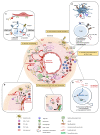
Summarized pathogenesis of Giant Cell Arterits (GCA). Step 1: an undefined danger signal activates vascular dendritic cells (DC) that then acquire a mature phenotype (CD83+CD80/86+CCR7+MHC-IIhigh) and produce chemokines (CCL18, CCL19, CCL20 and CCL21), leading to the recruitment of CCR6+CD161+CD4+ T cells. Step 2: CD4+ T cells are activated by DCs and polarize into Th1 and Th17 cells through the effect of IL-12, IL-23, IL-6 and IL-1β, which are produced by activated DC. Th1 and Th17 lymphocytes release IFN-γ and IL-17, respectively. Step 3: IFN-γ induces the activation of vascular smooth muscle cells (VSMC) in the media and enables them to produce chemokines (CCL2, CXCL9, CXCL10, CXCL11), which trigger the recruitment of additional T cells (CD4+ and CD8+) and monocytes. Monocytes differentiate into macrophages and merge into multinucleated giant cells, the hallmark of GCA. Step 4: vascular remodeling is characterized by the destruction of the internal elastic lamina and the proliferation and migration of VSMC into the intima. Macrophages play a key role in this process through the release of several factors such as Platelet-Derived Growth Factor (PDGF), reactive oxygen species (ROS), Matrix metalloproteinase-9 (MMP-9), IL-6, IL-1β, Granulocyte-Macrophage Colony-Stimulating Factor (GM-CSF) and TNF-α, which contribute to tissue damage and intimal hyperplasia. Likewise, VSMCs and endothelial cells release ET-1, which stimulates VSMC migration and proliferation and thus induces intimal hyperplasia. Moreover, macrophages and VSMCs also produce vascular endothelial growth factor (VEGF), which is responsible for neoangiogenesis and promotes a local inflammatory response. A cellular transition from VSMC to a myofibroblast phenotype is observed, and the accumulation of these cells in the neo-intima leads to vascular occlusion, which is responsible for ischemic complications of GCA. (AE) Mechanisms involved in the maintenance of vascular inflammation. (A): Programmed death-ligand 1 (PDL-1) defect on antigen-presenting cell surface leads to the persistent activity of T cells and contributes to hyperplasia and neoangiogenesis. (B): IL-6, which is a pro-inflammatory cytokine implicated in the GCA pathogenesis, impairs Tregs’ function, decreases their frequency and promotes the shift to a Treg deficiency in exon 2 of FoxP3 that is prone to producing IL-17. (C): In addition to their role in the recruitment of monocytes and vascular remodeling, VSMC differentiate into myofibroblasts, which also participate in the maintenance of Th1 and Tc1 polarizations. Moreover, myofibroblasts have an important capacity to produce extracellular matrix proteins (COL: collagen, FN: fibronectin) that contribute to the rigidification of the vascular wall. (D): GM-CSF is produced by CD4 T cells, macrophages, VSMC and endothelial cells. GM-CSF-Receptor-α is highly expressed in GCA lesions, and an autocrine amplification loop takes place. GM-CSF is involved in cell differentiation, vascular inflammation and vascular remodeling. (E): T cells and resident cells of the arterial wall, like endothelial cells, communicate via the NOTCH pathway. The ligation of Notch 1 to Jagged 1 decreases the polarization Th1/Th17. Otherwise, in CD8+Treg cells, aberrant Notch 4 signaling drives the suppression of RAB7A involved in the exosomal release of NOX2.
Figure 3
Figure 3
Therapeutic strategies in GCA. Steps 1–4 show the pathogenesis of the GCA physiopathology: 1—activation of dendritic cells; 2—activation, proliferation and polarization of T cells toward Th1 and Th17 cells; 3—effect of interferon-gamma (IFN-γ) on vascular smooth muscle cells (VSMCs) leading to the recruitment of additional CD4 T cells together with CD8 T cells and monocytes; 4—production of mediators implicated in vascular remodeling: neoangiogenesis (VEGF), migration, proliferation and differentiation of VSMCs into myofibroblasts, leading to hyperplastic neointima. The main drugs approved or under evaluation for the treatment of GCA are shown in the figure with their main therapeutic targets. Steroids inhibit T-cell activation, proliferation and polarization into Th17 cells. In addition, they trigger a decrease in the level of serum IL-6. Abatacept blocks T-cell activation through its ability to prevent interaction between CD28 and CD80/86. Methotrexate inhibits the proliferation of T cells and their ability to produce cytokines. Ustekinumab targets the p40 subunit, which is shared by IL-12 and IL-23. Guselkumab targets the p19 subunit of IL-23. Tocilizumab targets the receptors of IL-6. Mavrilimumab targets GM-CSF and should, therefore, impact vascular remodeling. Janus kinase inhibitors (JAKi) block the signaling pathways of several cytokines such as IL-6, IL-12 and IFN-γ and can thus theoretically inhibit the Th1 and Th17 pathways, to inhibit vascular inflammation and remodeling. DC: dendritic cells; VSMC: vascular smooth muscle cells; Mono: monocytes; MΦ: macrophages; PDGF: platelet-derived growth factor; VEGF: vascular endothelial growth factor; ET-1: endothelin-1; GM-CSF: granulocyte-macrophage colony-stimulating factor.

References

    1. Jennette J.C., Falk R.J., Bacon P.A., Basu N., Cid M.C., Ferrario F., Flores-Suarez L.F., Gross W.L., Guillevin L., Hagen E.C., et al. 2012 revised International Chapel Hill Consensus Conference Nomenclature of Vasculitides. Arthritis Rheum. 2013;65:1–11. doi: 10.1002/art.37715. - DOI - PubMed
    1. Espitia O., Samson M., Le Gallou T., Connault J., Landron C., Lavigne C., Belizna C., Magnant J., de Moreuil C., Roblot P., et al. Comparison of idiopathic (isolated) aortitis and giant cell arteritis-related aortitis. A French retrospective multicenter study of 117 patients. Autoimmun. Rev. 2016;15:571–576. doi: 10.1016/j.autrev.2016.02.016. - DOI - PubMed
    1. Berti A., Campochiaro C., Cavalli G., Pepe G., Praderio L., Sabbadini M.G., Dagna L. Giant cell arteritis restricted to the limb arteries: An overlooked clinical entity. Autoimmun. Rev. 2015;14:352–357. doi: 10.1016/j.autrev.2014.12.005. - DOI - PubMed
    1. Salvarani C., Cantini F., Hunder G.G. Polymyalgia rheumatica and giant-cell arteritis. Lancet. 2008;372:234–245. doi: 10.1016/S0140-6736(08)61077-6. - DOI - PubMed
    1. Salvarani C., Macchioni P., Zizzi F., Mantovani W., Rossi F., Castri C., Capozzoli N., Baricchi R., Boiardi L., Chiaravalloti F., et al. Epidemiologic and immunogenetic aspects of polymyalgia rheumatica and giant cell arteritis in northern Italy. Arthritis Rheum. 1991;34:351–356. doi: 10.1002/art.1780340313. - DOI - PubMed