Review
. 2022 May 22;11(10):2921.
doi: 10.3390/jcm11102921.

Transcatheter Treatment of Mitral Regurgitation

Affiliations
Review

Transcatheter Treatment of Mitral Regurgitation

Angela McInerney et al. J Clin Med. .

Abstract

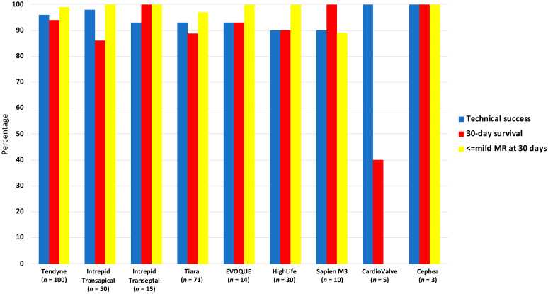
Mitral valve disease, and in particular mitral regurgitation, is a common clinical entity. Until recently, surgical repair and replacement were the only therapeutic options available, leaving many patients untreated mostly due to excessive surgical risk. Over the last number of years, huge strides have been made regarding percutaneous, catheter-based solutions for mitral valve disease. Transcatheter repair procedures have most commonly been used, and in recent years there has been exponential growth in the number of devices available for transcatheter mitral valve replacement. Furthermore, the evolution of these devices has resulted in both smaller delivery systems and a shift towards transeptal access, negating the need for surgical incisions. In line with these advancements, and clinical trials demonstrating promising outcomes in carefully selected cases, recent guidelines have strengthened their recommendations for these devices. It is appropriate, therefore, to now review the current transcatheter repair and replacement devices available and the evidence for their use.

Keywords: mitral valve; mitral valve repair; mitral valve replacement; transcatheter.


Conflict of interest statement

The authors declare no conflict of interest.

Figures

Figure 1
Figure 1
Transesophageal echocardiogram (TEE) pre-procedural assessment and intraprocedural guidance for a patient with severe secondary mitral regurgitation (MR) undergoing edge to edge repair. (A): mitral valve (MV) anatomy by 3D TEE showing the anterior leaflet (A) occupying one third of the annular perimeter and posterior leaflet (P) occupying two thirds. The anterior annulus has two fibrous trigones (T); the dashed red line represents the intertrigonal region. The commissures are marked with a green asterisk. The aortic valve sits anterior to the MV. Lateral (Lat) and Medial (Med) are also demarcated. (B,C) relationship between the MV and the coronary sinus (CS) and circumflex artery (LCx) on TEE; important for interventions targeting the annulus, the left atrial appendage (LAA) is seen laterally. Fossa height (FH) is demonstrated, which is important to determine feasibility of edge-to-edge repair procedures. (D): TTE view of the anterolateral papillary muscle (P) and its associated chordae (Ch). (EI): pre- and intra-procedural guidance by TEE for edge-to-edge repair of the MV in a patient with secondary MR. (E): Severe MR with a posteriorly directed regurgitant jet (effective regurgitant orifice area (EROA) 0.81 cm2). (F): 3D assessment of MV area (4.9 cm2 with mean transvalvular gradient of 3 mmHg). (G): Posterior leaflet length (length between blue asterisk) and fossa height (not shown in this image) are important to determine feasibility of edge-to-edge repair. (H): Intraprocedural transthoracic echocardiogram (TTE) showing the deployed clip (yellow asterisk) with the delivery catheter traversing the interatrial septum (red asterisk). (I): final result on 3D TEE with a double orifice MV TEE confirmed successful implantation with no significant stenosis (MV area 2 cm2, mean transvalvular gradient 5 mmHg) and mild residual MR.
Figure 2
Figure 2
TEE and cardiac computed tomography (CT) planning for a transcatheter Tendyne™ procedure in a patient with severe MR and calcified leaflets. (A): Severe MR with a posteriorly directed jet, (B): 3D transesophageal echo (TEE) demonstrating calcified leaflet tips unsuitable for edge-to-edge repair. (C): Calcified mitral valve (MV) leaflet tips, anterior MV leaflet (AMVL) length 25 mm (yellow) and AMVL to septum distance of 6 mm (red) (measured to assess risk of left ventricular outflow tract obstruction (LVOTO). (D): 3D assessment of MV annular area (A1 = 12 cm2), perimeter (13.1 cm), AP dimension (D2 = 3.11 cm), inter-trigonal distance (D1 = 3 cm) and intercommisural distance (D3 = 4.6 cm). (E): CT planning demonstrating MV dimensions (T to T = intertrigonal distance, perimeter outlined in red, anteroposterior (AP) distance and intercommisural distance (C to C)). (F): Simulated Tendyne™ with predicted neo-LVOT diameter (yellow asterisk). (G): Simulated Tendyne with predicted neo-left ventricular outflow tract (neo-LVOT) area of 4.6 cm2 (white shaded area), (H): CT assessed apical puncture site for correct orientation with the MV. (I): Intraprocedural TEE guidance with X-plane views showing the delivery system across the MV and in the left atrium (LA), (J): Partial liberation of the Tendyne™ system (LP37M) and assessment of paravalvular leak with colour Doppler. (K): Liberated Tendyne™ valve with no residual MR, no paravalvular leak, mean transvalvular gradient of 5 mmHg, and no dynamic gradient in the LVOT.
Figure 3
Figure 3
Transcatheter mitral valve repair systems.
Figure 4
Figure 4
Transcatheter Mitral Valve Replacement.
Figure 5
Figure 5
Procedural results following transcatheter mitral valve replacement.

References

    1. Iung B., Delgado V., Rosenhek R., Price S., Prendergast B., Wendler O., De Bonis M., Tribouilloy C., Evangelista A., Bogachev-Prokophiev A., et al. Contemporary Presentation and Management of Valvular Heart Disease: The EURObservational Research Programme Valvular Heart Disease II Survey. Circulation. 2019;140:1156–1169. doi: 10.1161/CIRCULATIONAHA.119.041080. - DOI - PubMed
    1. Cahill T.J., Prothero A., Wilson J., Kennedy A., Brubert J., Masters M., Newton J.D., Dawkins S., Enriquez-Sarano M., Prendergast B.D., et al. Community prevalence, mechanisms and outcome of mitral or tricuspid regurgitation. Heart. 2021;107:1003–1009. doi: 10.1136/heartjnl-2020-318482. - DOI - PubMed
    1. Vahanian A., Beyersdorf F., Praz F., Milojevic M., Baldus S., Bauersachs J., Capodanno D., Conradi L., De Bonis M., De Paulis R., et al. 2021 ESC/EACTS Guidelines for the management of valvular heart disease. Eur. Heart J. 2021;43:561–632. doi: 10.1093/eurheartj/ehab395. - DOI - PubMed
    1. Otto C.M., Nishimura R.A., Bonow R.O., Carabello B.A., Erwin J.P., Gentile F., 3rd, Jneid H., Krieger E.V., Mack M., McLeod C., et al. 2020 ACC/AHA Guideline for the Management of Patients with Valvular Heart Disease: A Report of the American College of Cardiology/American Heart Association Joint Committee on Clinical Practice Guidelines. Circulation. 2021;143:e72–e227. - PubMed
    1. Mack M., Carroll J.D., Thourani V., Vemulapalli S., Squiers J., Manandhar P., Deeb G.M., Batchelor W., Herrmann H.C., Cohen D.J., et al. Transcatheter Mitral Valve Therapy in the United States: A Report From the STS-ACC TVT Registry. J. Am. Coll. Cardiol. 2021;78:2326–2353. doi: 10.1016/j.jacc.2021.07.058. - DOI - PubMed

LinkOut - more resources