. 2022 Apr 30;12(5):727.
doi: 10.3390/jpm12050727.

Validation of a Novel Three-Dimensional (3D Fusion) Gross Sampling Protocol for Clear Cell Renal Cell Carcinoma to Overcome Intratumoral Heterogeneity: The Meet-Uro 18 Study

Matteo Brunelli  1   2 Guido Martignoni  1   3 Giorgio Malpeli  4 Alessandro Volpe  5 Luca Cima  6 Maria Rosaria Raspollini  7 Mattia Barbareschi  6 Alessandro Tafuri  8 Giulia Masi  9 Luisa Barzon  9 Serena Ammendola  1 Manuela Villanova  1 Maria Angela Cerruto  8 Michele Milella  10 Sebastiano Buti  11 Melissa Bersanelli  11 Giuseppe Fornarini  12 Sara Elena Rebuzzi  12 Valerio Gaetano Vellone  13 Gabriele Gaggero  13 Giuseppe Procopio  14 Elena Verzoni  14 Sergio Bracarda  15 Martina Fanelli  16 Roberto Sabbatini  16 Rodolfo Passalacqua  17 Bruno Perrucci  17 Maria Olga Giganti  17 Maddalena Donini  17 Stefano Panni  17 Marcello Tucci  18 Veronica Prati  19 Cinzia Ortega  19 Anna Caliò  1   2 Albino Eccher  1 Filippo Alongi  20 Giovanni Pappagallo  21 Roberto Iacovelli  22 Alessandra Mosca  23 Paolo Umari  7 Ilaria Montagnani  7 Stefano Gobbo  24 Francesco Atzori  25 Enrico Munari  26 Marco Maruzzo  27 Umberto Basso  27 Francesco Pierconti  28 Carlo Patriarca  29 Piergiuseppe Colombo  30 Alberto Lapini  31 Giario Conti  32 Roberto Salvioni  33 Enrico Bollito  34 Andrea Cossarizza  35 Francesco Massari  36 Mimma Rizzo  37 Renato Franco  38 Federica Zito-Marino  38 Yoseba Aberasturi Plata  39 Francesca Galuppini  40 Marta Sbaraglia  40 Matteo Fassan  40 Angelo Paolo Dei Tos  40 Maurizio Colecchia  41 Holger Moch  42 Maurizio Scaltriti  43 Camillo Porta  44   45 Brett Delahunt  46 Gianluca Giannarini  47 Roberto Bortolus  48 Pasquale Rescigno  49 Giuseppe Luigi Banna  49 Alessio Signori  50 Miguel Angel Llaja Obispo  51 Roberto Perris  52 Alessandro Antonelli  8
Affiliations

Validation of a Novel Three-Dimensional (3D Fusion) Gross Sampling Protocol for Clear Cell Renal Cell Carcinoma to Overcome Intratumoral Heterogeneity: The Meet-Uro 18 Study

Matteo Brunelli et al. J Pers Med. .

Abstract

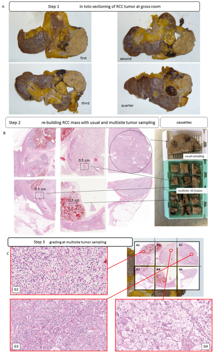
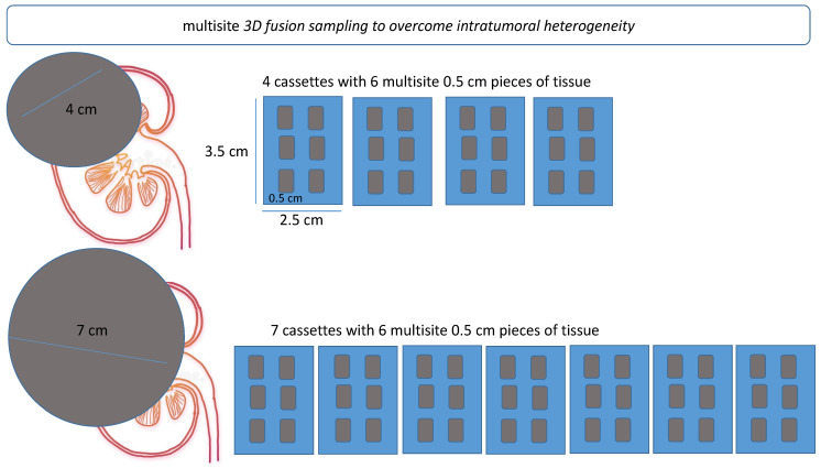
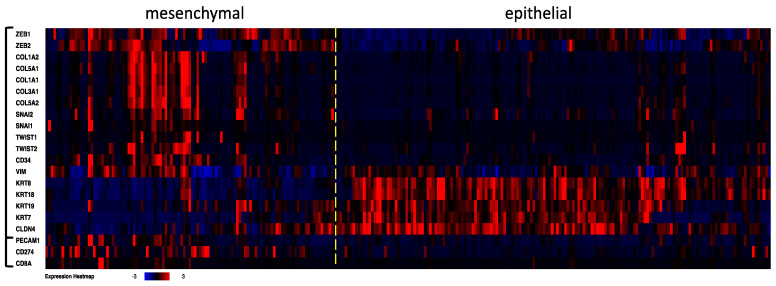
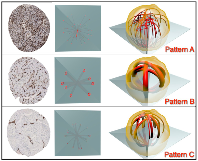
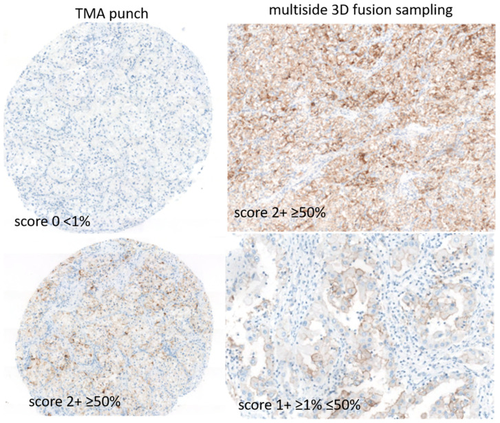
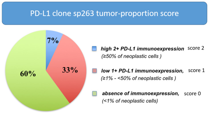
We aimed to overcome intratumoral heterogeneity in clear cell renal cell carcinoma (clearRCC). One hundred cases of clearRCC were sampled. First, usual standard sampling was applied (1 block/cm of tumor); second, the whole tumor was sampled, and 0.6 mm cores were taken from each block to construct a tissue microarray; third, the residual tissue, mapped by taking pieces 0.5 × 0.5 cm, reconstructed the entire tumor mass. Precisely, six randomly derived pieces of tissues were placed in each cassette, with the number of cassettes being based on the diameter of the tumor (called multisite 3D fusion). Angiogenic and immune markers were tested. Routine 5231 tissue blocks were obtained. Multisite 3D fusion sections showed pattern A, homogeneous high vascular density (10%), pattern B, homogeneous low vascular density (8%) and pattern C, heterogeneous angiogenic signatures (82%). PD-L1 expression was seen as diffuse (7%), low (33%) and absent (60%). Tumor-infiltrating CD8 scored high in 25% (pattern hot), low in 65% (pattern weak) and zero in 10% of cases (pattern desert). Grading was upgraded in 26% of cases (G3-G4), necrosis and sarcomatoid/rhabdoid characters were observed in, respectively, 11 and 7% of cases after 3D fusion (p = 0.03). CD8 and PD-L1 immune expressions were higher in the undifferentiated G4/rhabdoid/sarcomatoid clearRCC subtypes (p = 0.03). Again, 22% of cases were set to intermediate to high risk of clinical recurrence due to new morphological findings of all aggressive G4, sarcomatoid/rhabdoid features by using 3D fusion compared to standard methods (p = 0.04). In conclusion, we propose an easy-to-apply multisite 3D fusion sampling that negates bias due to tumor heterogeneity.

Keywords: angiogenesis; clear cell renal cell carcinoma; immunity; immunohistochemistry; intratumoral heterogeneity; tumor sampling.


Conflict of interest statement

The authors declare no conflict of interest.

Figures

Figure 1
Figure 1
Intratumoral heterogeneity in clear cell RCCs. Gross images of different slices of clear cell RCCs with heterogenous macroscopic appearance (A); usual block (white cassette) and multisite sampling with 6 pieces of tumor tissue per single block (green cassette) and 3D fusion multisite reconstruction (B,C); H&E staining (C).
Figure 2
Figure 2
Schematic representation of multisite 3D fusion sampling procedure. Fusion sampling was based on the size of the tumor. For each cm diameter of tumor, six blocks of tissue, each measuring 0.5 × 0.5 cm, were randomly selected and placed in an individual cassette.
Figure 3
Figure 3
Transcriptome investigation in 512 clear cell RCC cases from TCGA (PanCancer Atlas study). The mRNA levels are z-scores relative to diploid samples (RNA Seq V2 RSEM). Clusterization procedure was applied to the first 18 genes, then the PECAM1 (CD31), CD274 (PD-L1) and CD8A mRNA levels were added to the established case list order. A dashed line was added to separate mesenchymal and epithelial clearRCC cases. The heat map shows angiogenesis and immune signatures in the clearRCC epithelial and mesenchymal subtypes. Higher levels of angiogenic markers CD31 and CD34 and of the immune markers PD-L1 and CD8A come up predominantly in the mesenchymal subtype.
Figure 4
Figure 4
Classification of angiogenesis in clear cell RCCs after 3D fusion multisite tumor sampling. Diagram showing categorization of pattern of angiogenesis: pattern A, characterized by homogeneous high level of angiogenesis; pattern B, characterized by homogeneous low level of angiogenesis; and pattern C, characterized by a mixture of patterns.
Figure 5
Figure 5
Classification of PD-L1 immune activation in clear cell RCCs after 3D fusion multisite tumor sampling. Diagram showing categorization of immune patterns: score 2 strong expression; score 1, low/weak expression; score 0 with absence of any expression.
Figure 6
Figure 6
Distribution of PD-L1 immunohistochemical expression in clear cell RCCs after 3D fusion multisite tumor sampling.

References

    1. López J.I. Intratumor heterogeneity in clear cell renal cell carcinoma: A review for the practicing pathologist. APMIS. 2016;124:153–159. doi: 10.1111/apm.12500. - DOI - PubMed
    1. Longo D.L. Tumor Heterogeneity and Personalized Medicine. N. Engl. J. Med. 2012;366:956–957. doi: 10.1056/NEJMe1200656. - DOI - PubMed
    1. McGranahan N., Burrell R.A., Endesfelder D., Novelli M.R., Swanton C. Cancer chromosomal instability: Therapeutic and diagnostic challenges. EMBO Rep. 2012;13:528–538. doi: 10.1038/embor.2012.61. - DOI - PMC - PubMed
    1. Gerlinger M., Horswell S., Larkin J., Rowan A.J., Salm M., Varela I., Fisher R., McGranahan N., Matthews N., Santos C.R., et al. Genomic architecture and evolution of clear cell renal cell carcinomas defined by multiregion sequencing. Nat. Genet. 2014;46:225–233. doi: 10.1038/ng.2891. - DOI - PMC - PubMed
    1. Msaouel P., Malouf G.G., Su X., Yao H., Tripathi D., Soeung M., Gao J., Rao P., Coarfa C., Creighton C.J., et al. Comprehensive Molecular Characterization Identifies Distinct Genomic and Immune Hallmarks of Renal Medullary Carcinoma. Cancer Cell. 2020;37:720–734.e13. doi: 10.1016/j.ccell.2020.04.002. - DOI - PMC - PubMed