Review
. 2022 May 5;12(5):747.
doi: 10.3390/jpm12050747.

Targeting HIF-1α Function in Cancer through the Chaperone Action of NQO1: Implications of Genetic Diversity of NQO1

Affiliations
Review

Targeting HIF-1α Function in Cancer through the Chaperone Action of NQO1: Implications of Genetic Diversity of NQO1

Eduardo Salido et al. J Pers Med. .

Abstract

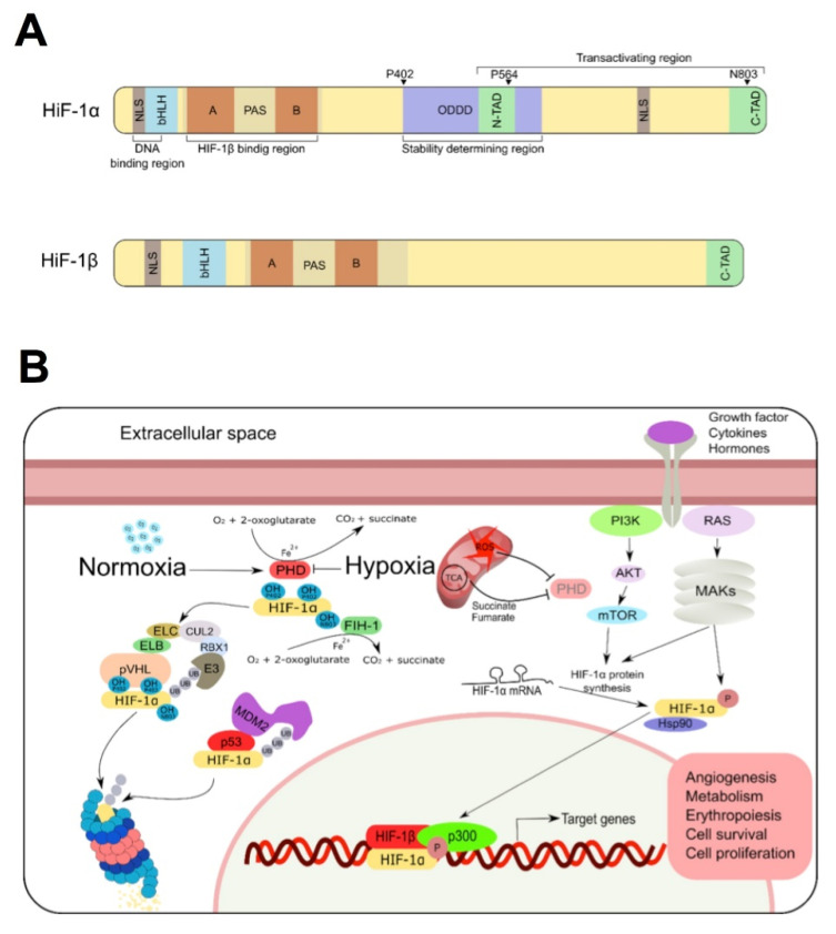
HIF-1α is a master regulator of oxygen homeostasis involved in different stages of cancer development. Thus, HIF-1α inhibition represents an interesting target for anti-cancer therapy. It was recently shown that the HIF-1α interaction with NQO1 inhibits proteasomal degradation of the former, thus suggesting that targeting the stability and/or function of NQO1 could lead to the destabilization of HIF-1α as a therapeutic approach. Since the molecular interactions of NQO1 with HIF-1α are beginning to be unraveled, in this review we discuss: (1) Structure-function relationships of HIF-1α; (2) our current knowledge on the intracellular functions and stability of NQO1; (3) the pharmacological modulation of NQO1 by small ligands regarding function and stability; (4) the potential effects of genetic variability of NQO1 in HIF-1α levels and function; (5) the molecular determinants of NQO1 as a chaperone of many different proteins including cancer-associated factors such as HIF-1α, p53 and p73α. This knowledge is then further discussed in the context of potentially targeting the intracellular stability of HIF-1α by acting on its chaperone, NQO1. This could result in novel anti-cancer therapies, always considering that the substantial genetic variability in NQO1 would likely result in different phenotypic responses among individuals.

Keywords: HIF-1α; NQO1; angiogenesis; cancer; genetic variability; hypoxia; ligand binding; proteasomal degradation; protein: protein interactions.


Conflict of interest statement

The authors declare no conflict of interest. The funders had no role in the design of the study; in the collection, analyses, or interpretation of data; in the writing of the manuscript, or in the decision to publish the results.

Figures

Figure 3
Figure 3
Structure and catalytic function of NQO1. (A) Structural representation of the NQO1 dimer (PDB 2F1O) [83]. NTD and CTD refer to N-terminal and C-terminal domains, respectively. The location of the FAD and the inhibitor dicoumarol (DIC) binding sites is also indicated. The monomer:monomer interface is indicated as MMI. (B) Reduction of FAD by NADH shows two different pathways. In the left panel, the spectral properties of the different spectroscopic species (A, B, C) stabilized upon reduction are indicated as well as their conversion limiting rate constants. The right panel shows the model proposed by us for the sequential reduction of the two FAD cofactors in the protein homo-dimer, in which this large difference in kinetics represents a type of functional negative cooperativity. Adapted from [76].
Figure 4
Figure 4
The NQO1 interactome. (A) NQO1 protein partners described in at least one report. (B) Connectivity between NQO1 partners. The shorter the radial distance between a partner and NQO1 indicates a higher interconnectivity. Partners in yellow are from mouse and in blue from human. (C) A zoom from panel (B) shows the most highly interconnected partners of NQO1. (DJ) Interactions between NQO1 partners in panel (C) and other proteins to highlight their different interconnectivity. The thickness of connecting yellow lines is related to the number of reports describing the interaction. Data were retrieved from the BioGRID database (https://thebiogrid.org/108072, accessed on 1 April 2022) [111].
Figure 1
Figure 1
Structure and regulation of HIF. (A) Schematic representation of human HIF-1α and HIF-1β structure. Both proteins form part of the HLH–PAS transcription factor family and contain a N-terminal bHLH domain (implicated in DNA binding) and two PAS domains (responsible of its dimerization). HIF-1α contains an oxygen-dependent degradation domain (ODDD) that mediates oxygen-regulated stability, and two transactivation domains (TAD), that mediate its transcriptional activity and its stability. (B) In normoxia, HIF-1α is subjected to oxygen-dependent hydroxylation by Prolyl hydroxylase domain (PHD) hydroxylases, conducting HIF-1α to ubiquitination by the von Hippel-Lindau protein (pVHL) and proteasomal degradation. MDM2/p53 are also involved in ubiquitination and degradation of HIF1α protein in a pVHL-independent manner. Under low oxygen levels (as well as some metabolites of TCA cycle), PHD is inhibited, HIF-1α translocates to the nucleus and promotes the activation of target genes. Transactivation of HIF-1α could be also induced by PI3K/MAPKs signaling pathways upon activation of some growth factor, hormones and cytokines.
Figure 2
Figure 2
Nuclear HIF-1α (top) and NQO1 (bottom) expression (brown staining) in serial sections of clear cell renal carcinoma (ccRCC; this particular case carried p.L89L mutation in one allele and a deletion of the other one). Common deletions of the VHL gene result in lack of HIF1 degradation, with the subsequent activation of HIF1 as a nuclear transcription factor that drives tumor cell proliferation. NQO1 is widely expressed in the cytoplasm of tumor cells, including clear cell renal carcinoma, and it might play a role in the postranslational regulation of HIF-1α. Clear cell ovarian carcinoma is another example with strong hypoxic signature where NQO1 might play a role, however we have had no access to cases of clear cell ovarian carcinoma driven by ARID1A mutations. Much more work is needed to address this interesting connection. Primary antibodies used: rabbit polyclonal anti-HIF1α (ab2185, dilution 1:100, Abcam, Cambridge, UK) and mouse monoclonal anti-NQO1 (clone A180, dilution 1:600, Thermofisher scientific, Madrid, Spain); peroxidase signal development with Optiview, Ventana.
Figure 2
Figure 2
Nuclear HIF-1α (top) and NQO1 (bottom) expression (brown staining) in serial sections of clear cell renal carcinoma (ccRCC; this particular case carried p.L89L mutation in one allele and a deletion of the other one). Common deletions of the VHL gene result in lack of HIF1 degradation, with the subsequent activation of HIF1 as a nuclear transcription factor that drives tumor cell proliferation. NQO1 is widely expressed in the cytoplasm of tumor cells, including clear cell renal carcinoma, and it might play a role in the postranslational regulation of HIF-1α. Clear cell ovarian carcinoma is another example with strong hypoxic signature where NQO1 might play a role, however we have had no access to cases of clear cell ovarian carcinoma driven by ARID1A mutations. Much more work is needed to address this interesting connection. Primary antibodies used: rabbit polyclonal anti-HIF1α (ab2185, dilution 1:100, Abcam, Cambridge, UK) and mouse monoclonal anti-NQO1 (clone A180, dilution 1:600, Thermofisher scientific, Madrid, Spain); peroxidase signal development with Optiview, Ventana.
Figure 5
Figure 5
Subcellular location of NQO1 interacting human partners based on data from GeneCards® (https://www.genecards.org/, accessed on 1 April 2022). For all the interactors, the subcellular compartment (location) and its confidence (1–5, from the lowest to the highest) were retrieved from GeneCards®. (A) Accumulated score for each organelle as the sum of the numerical degree of confidence for all partners found in a given compartment. The highest accumulated score (i.e., for the nucleus) was used to normalize yielding the Score. As Fraction, we refer to the fraction of all the partners found in a given organelle. Note that the ratio Score:Fraction gives a measure of the degree of confidence for finding a given partner in a given subcellular location. (B) Subcellular location of NQO1 partners in the three subcellular locations of NQO1 reported with confidence (i.e., equal or higher than 3) as the fraction of the total of NQO1 partners. Overlapping regions in the Venn diagram reveal the presence of an NQO1 partner in at least two subcellular locations. Numbers in the different regions of the Venn diagram represent the fraction of NQO1 partners present in different subcellular locations.
Figure 6
Figure 6
Structure and dynamics of NQO1 upon binding different ligands. (A) Structural overlay of the X-ray structures of NQO1holo (1D4A) [81], NQO1holo:NAD+ (kindly supplied by Profs. Mario Bianchet and Mario Amzel, John Hopkins University Medical School, Baltimore, Maryland, USA) and NQO1dic (2F1O). The lower panel shows a zoom highlighting the position of the FAD (orange, NQO1dic and red, NQO1holo:NAD+), NAD+ (in grey) and dicoumarol (Dic, in black). (B) Dicoumarol binding causes long-range effects on the structural dynamics of NQO1 WT. Residues shown in dot representation are those for which the structural dynamics is reduced according to HDXMS [132]. (C) Most of the residues whose dynamics are reduced upon dicoumarol binding are solvent-exposed. The plot in the left shows the solvent accessible surface area (SASA) for the each residue as calculated in [132] and color circles indicate the magnitude of the change in structural dynamics. The figure on the right shows the structural location of solvent-exposed residues (SASA > 20%). The color scales in panels B and C reflect the magnitude of the changes in protein dynamics according to [132] and red arrows indicate the position of dicoumarol.

References

    1. Semenza G.L. Hypoxia-inducible factors in physiology and medicine. Cell. 2012;148:399–408. doi: 10.1016/j.cell.2012.01.021. - DOI - PMC - PubMed
    1. Nakazawa M.S., Keith B., Simon M.C. Oxygen availability and metabolic adaptations. Nat. Rev. Cancer. 2016;16:663–673. doi: 10.1038/nrc.2016.84. - DOI - PMC - PubMed
    1. Keith B., Johnson R.S., Simon M.C. HIF-1α and HIF-2α: Sibling rivalry in hypoxic tumour growth and progression. Nat. Rev. Cancer. 2012;12:9–22. doi: 10.1038/nrc3183. - DOI - PMC - PubMed
    1. Wang G.L., Jiang B.H., Rue E.A., Semenza G.L. Hypoxia-inducible factor 1 is a basic-helix-loop-helix-PAS heterodimer regulated by cellular O2 tension. Proc. Natl. Acad. Sci. USA. 1995;92:5510–5514. doi: 10.1073/pnas.92.12.5510. - DOI - PMC - PubMed
    1. Yee Koh M., Spivak-Kroizman T.R., Powis G. HIF-1 regulation: Not so easy come, easy go. Trends Biochem. Sci. 2008;33:526–534. doi: 10.1016/j.tibs.2008.08.002. - DOI - PubMed