

An Integrated Approach on the Diagnosis of Cerebral Veins and Dural Sinuses Thrombosis (a Narrative Review)
- PMID: 35629384
- PMCID: PMC9145675
- DOI: 10.3390/life12050717
An Integrated Approach on the Diagnosis of Cerebral Veins and Dural Sinuses Thrombosis (a Narrative Review)
Abstract
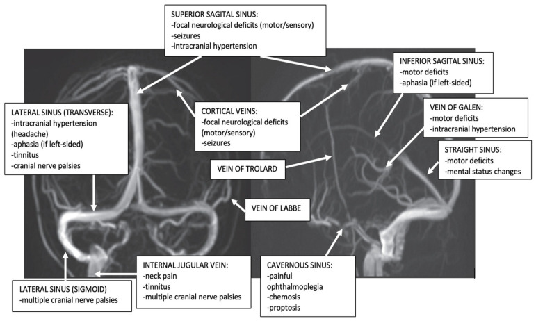
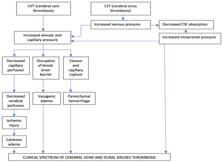
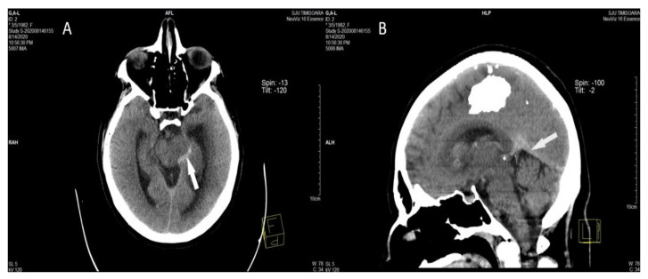
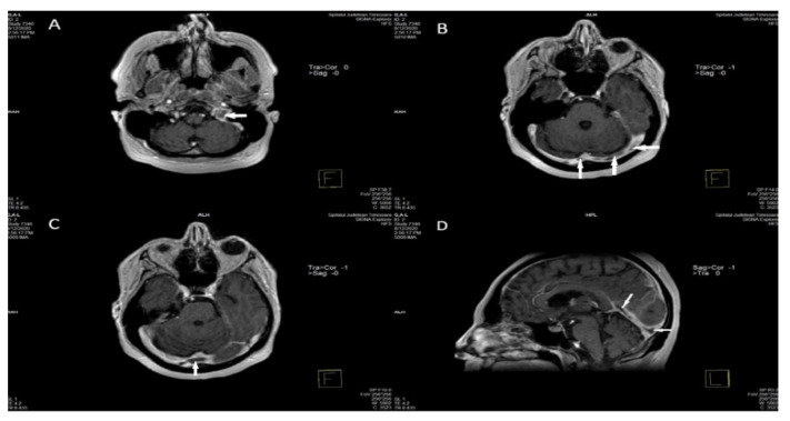
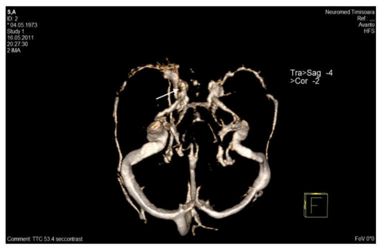
(1) Objective: This review paper aims to discuss multiple aspects of cerebral venous thrombosis (CVT), including epidemiology, etiology, pathophysiology, and clinical presentation. Different neuroimaging methods for diagnosis of CVT, such as computer tomography CT/CT Venography (CTV), and Magnetic Resonance Imaging (MRI)/MR Venography (MRV) will be presented. (2) Methods: A literature analysis using PubMed and the MEDLINE sub-engine was done using the terms: cerebral venous thrombosis, thrombophilia, and imaging. Different studies concerning risk factors, clinical picture, and imaging signs of patients with CVT were examined. (3) Results: At least one risk factor can be identified in 85% of CVT cases. Searching for a thrombophilic state should be realized for patients with CVT who present a high pretest probability of severe thrombophilia. Two pathophysiological mechanisms contribute to their highly variable clinical presentation: augmentation of venular and capillary pressure, and diminution of cerebrospinal fluid absorption. The clinical spectrum of CVT is frequently non-specific and presents a high level of clinical suspicion. Four major syndromes have been described: isolated intracranial hypertension, seizures, focal neurological abnormalities, and encephalopathy. Cavernous sinus thrombosis is the single CVT that presents a characteristic clinical syndrome. Non-enhanced CT (NECT) of the Head is the most frequently performed imaging study in the emergency department. Features of CVT on NECT can be divided into direct signs (demonstration of dense venous clot within a cerebral vein or a cerebral venous sinus), and more frequently indirect signs (such as cerebral edema, or cerebral venous infarct). CVT diagnosis is confirmed with CTV, directly detecting the venous clot as a filling defect, or MRI/MRV, which also realizes a better description of parenchymal abnormalities. (4) Conclusions: CVT is a relatively rare disorder in the general population and is frequently misdiagnosed upon initial examination. The knowledge of wide clinical aspects and imaging signs will be essential in providing a timely diagnosis.
Keywords: Magnetic Resonance (MR) Venography; Magnetic Resonance Imaging (MRI) of the Head; cerebral veins and dural sinuses thrombosis (CVT); headache; native and contrast-enhanced Head Computed Tomography (CT); thrombophilia.
Conflict of interest statement
The authors declare no competing interest.
Figures
References
-
- Bousser M.G., Barnett H.J.M. Chapter Twelve: Cerebral Venous Thrombosis. In: Mohr J.P., Choi D.W., Grotta J.C., Weir B., Wolf P.A., editors. Stroke (Pathophysiology, Diagnosis, and Management) 4th ed. Churchill Livingstone; London, UK: 2004. pp. 301–325.
-
- Ferro J.M., Canhão P. Chapter 45: Cerebral Venous Thrombosis. In: Grotta J.C., Albers G.W., Broderick J.P., Kasner S.E., Lo E.H., Mendelow A.D., Sacco R.L., Wong L.K.S., editors. Stroke (Pathophysiology, Diagnosis, and Management) 6th ed. Elsevier; Amsterdam, The Netherlands: 2016. pp. 716–730.
Publication types
LinkOut - more resources
Full Text Sources
