Review
. 2022 May 3;11(5):538.
doi: 10.3390/pathogens11050538.

The Evolutionary Dance between Innate Host Antiviral Pathways and SARS-CoV-2

Affiliations
Review

The Evolutionary Dance between Innate Host Antiviral Pathways and SARS-CoV-2

Saba R Aliyari et al. Pathogens. .

Abstract

Compared to what we knew at the start of the SARS-CoV-2 global pandemic, our understanding of the interplay between the interferon signaling pathway and SARS-CoV-2 infection has dramatically increased. Innate antiviral strategies range from the direct inhibition of viral components to reprograming the host's own metabolic pathways to block viral infection. SARS-CoV-2 has also evolved to exploit diverse tactics to overcome immune barriers and successfully infect host cells. Herein, we review the current knowledge of the innate immune signaling pathways triggered by SARS-CoV-2 with a focus on the type I interferon response, as well as the mechanisms by which SARS-CoV-2 impairs those defenses.

Keywords: COVID-19; SARS-CoV-2; innate immunity; interferon.


Conflict of interest statement

The authors declare no conflict of interest.

Figures
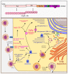
Figure 1
Figure 1
Genome organization and life cycle of SARS-CoV-2. Upper box: The SARS-CoV-2 genome consists of a 5′-cap, ORF1ab (encodes nsp1-16), spike protein, ORF3 (encodes ORF3a protein), E (envelope protein), M (membrane glycoprotein), ORF6 (ORF6 protein), ORF7 (ORF7a and ORF7b proteins), ORF8 (ORF8 protein), N (nucleocapsid), ORF10 (ORF10 protein), and a 3′-polyA tail. Lower box: SARS-CoV-2 binds the ACE2 receptor and fuses with the cell membrane. The plus-sense viral RNA genome is released and directly translated into polyproteins 1a and 1ab. These are cleaved to form the nsp proteins and RNA-dependent RNA polymerase (RdRp). RdRp produces the genome, anti-genome, and sub-genome copies of mRNA that encode the remaining viral proteins, including the nucleocapsid (N), membrane (M), envelope (E), and spike (S) proteins. N assembles with the genome copies while the other proteins are modified in the ER–Golgi-intermediate compartment (ERGIC), which then come together to form a mature virion that is released by exocytosis.
Figure 2
Figure 2
Taxonomy of the Coronaviridae family.Coronaviridae is divided into four groups of viruses: alphacoronaviruses include hCoV-229E and NL-63. The betacoronavirus group includes three subgroups: the Embecoviruses with hCoV-HKU-1 and OC43, the Sarbecoviruses with SARS-CoV-1 and SARS-CoV-2, and the Merbecovirus MERS-CoV.
Figure 3
Figure 3
Innate immune signaling pathways activated during coronavirus infection. Viral RNA stimulation of the RIG-I/MDA5 pathway activates the downstream IRF3 and NF-κB pathways to induce IFN-I production. IFN-I members (such as β) then bind the IFNAR1 receptor and trigger the Jak/Stat pathway and the expression of ISGs in neighboring cells [20]. Coronavirus proteins have evolved ways to target components of these signaling pathways to prevent the interferon response. PLpro, nsp13, and nsp16 interfere with MDA5 activation. PLpro, nsp6, N, nsp12, ORF3b, and ORF6 interfere with IRF3 activation. NF-κB activation is inhibited by nsp13 and ORF9c, and Jak/Stat activation is suppressed by ORF3a. Coronavirus proteins also interfere with host cellular processes; for example, nsp1, nsp10, nsp14, and N block translation, and ORF5 blocks nuclear trafficking.
Figure 4
Figure 4
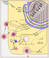
The ISG response triggers multiple mechanisms to combat coronavirus infection. ISGs interfere with viral entry at the plasma membrane and endocytosis (CH25H, IFITM, Ly6E), genome release (ZAP, OAS), and budding (activation of Bst2, inhibition of FASN).

References

    1. Li F. Structure, Function, and Evolution of Coronavirus Spike Proteins. Annu. Rev. Virol. 2016;3:237–261. doi: 10.1146/annurev-virology-110615-042301. - DOI - PMC - PubMed
    1. Tang Q., Song Y., Shi M., Cheng Y., Zhang W., Xia X.Q. Inferring the hosts of coronavirus using dual statistical models based on nucleotide composition. Sci. Rep. 2015;5:17155. doi: 10.1038/srep17155. - DOI - PMC - PubMed
    1. Megger D.A., Philipp J., Le-Trilling V.T.K., Sitek B., Trilling M. Deciphering of the Human Interferon-Regulated Proteome by Mass Spectrometry-Based Quantitative Analysis Reveals Extent and Dynamics of Protein Induction and Repression. Front. Immunol. 2017;8:1139. doi: 10.3389/fimmu.2017.01139. - DOI - PMC - PubMed
    1. Akira S. Pathogen recognition by innate immunity and its signaling. Proc. Jpn. Acad. Ser. B Phys. Biol. Sci. 2009;85:143–156. doi: 10.2183/pjab.85.143. - DOI - PMC - PubMed
    1. Fensterl V., Sen G.C. Interferons and viral infections. BioFactors. 2009;35:14–20. doi: 10.1002/biof.6. - DOI - PubMed