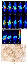
An Albumin-Binding PSMA Ligand with Higher Tumor Accumulation for PET Imaging of Prostate Cancer
- PMID: 35631340
- PMCID: PMC9143078
- DOI: 10.3390/ph15050513
An Albumin-Binding PSMA Ligand with Higher Tumor Accumulation for PET Imaging of Prostate Cancer
Abstract
Prostate-specific membrane antigen (PSMA) is an ideal target for the diagnosis and treatment of prostate cancer. Due to the short half-life in blood, small molecules/peptides are rapidly cleared by the circulatory system. Prolonging the half-life of PSMA probes has been considered as an effective strategy to improve the tumor detection. Herein, we reported a 64Cu-labeled PSMA tracer conjugating with maleimidopropionic acid (MPA), 64Cu-PSMA-CM, which showed an excellent ability to detect PSMA-overexpressing tumors in delayed time. Cell experiments in PSMA-positive 22Rv1 cells, human serum albumin binding affinity, and micro-PET imaging studies in 22Rv1 model were performed to investigate the albumin binding capacity and PSMA specificity. Comparisons with 64Cu-PSMA-BCH were performed to explore the influence of MPA on the biological properties. 64Cu-PSMA-CM could be quickly prepared within 30 min. The uptake of 64Cu-PSMA-CM in 22Rv1 cells increased over time and it could bind to HSA with a high protein binding ratio (67.8 ± 1.5%). When compared to 64Cu-PSMA-BCH, 64Cu-PSMA-CM demonstrated higher and prolonged accumulation in 22Rv1 tumors, contributing to high tumor-to-organ ratios. These results showed that 64Cu-PSMA-CM was PSMA specific with a higher tumor uptake, which demonstrated that MPA is an optional strategy for improving the radioactivity concentration in PSMA-expressing tumors and for developing the ligands for PSMA radioligand therapy.
Keywords: PET imaging; PSMA; copper radioisotopes; maleimidopropionic acid; prostate cancer.
Conflict of interest statement
The authors declare no conflict of interest.
Figures


References
-
- Okarvi S.M. Recent developments of prostate-specific membrane antigen (PSMA)-specific radiopharmaceuticals for precise imaging and therapy of prostate cancer: An overview. Clin. Transl. Imaging. 2019;7:189–208. doi: 10.1007/s40336-019-00326-3. - DOI
-
- Hofman M.S., Emmett L., Sandhu S., Iravani A., Joshua A.M., Goh J.C., Pattison D.A., Tan T.H., Kirkwood I.D., Ng S., et al. [177Lu]Lu-PSMA-617 versus cabazitaxel in patients with metastatic castration-resistant prostate cancer (TheraP): A randomised, open-label, phase 2 trial. Lancet. 2021;397:797–804. doi: 10.1016/S0140-6736(21)00237-3. - DOI - PubMed
-
- Zacho H.D., Ravn S., Afshar-Oromieh A., Fledelius J., Ejlersen J.A., Petersen L.J. Added value of (68)Ga-PSMA PET/CT for the detection of bone metastases in patients with newly diagnosed prostate cancer and a previous (99m)Tc bone scintigraphy. EJNMMI Res. 2020;10:31. doi: 10.1186/s13550-020-00618-0. - DOI - PMC - PubMed
Grants and funding
LinkOut - more resources
Full Text Sources
Miscellaneous

