Review
. 2022 May 23;14(10):2123.
doi: 10.3390/polym14102123.

Bone Mineralization in Electrospun-Based Bone Tissue Engineering

Affiliations
Review

Bone Mineralization in Electrospun-Based Bone Tissue Engineering

Dong-Jin Lim. Polymers (Basel). .

Abstract

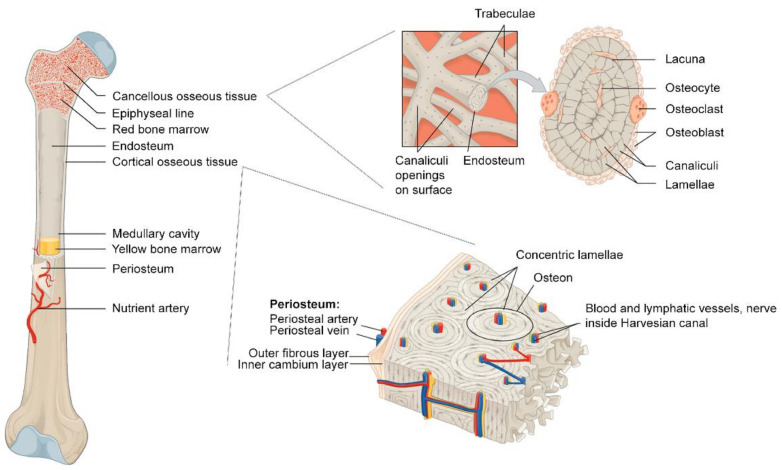
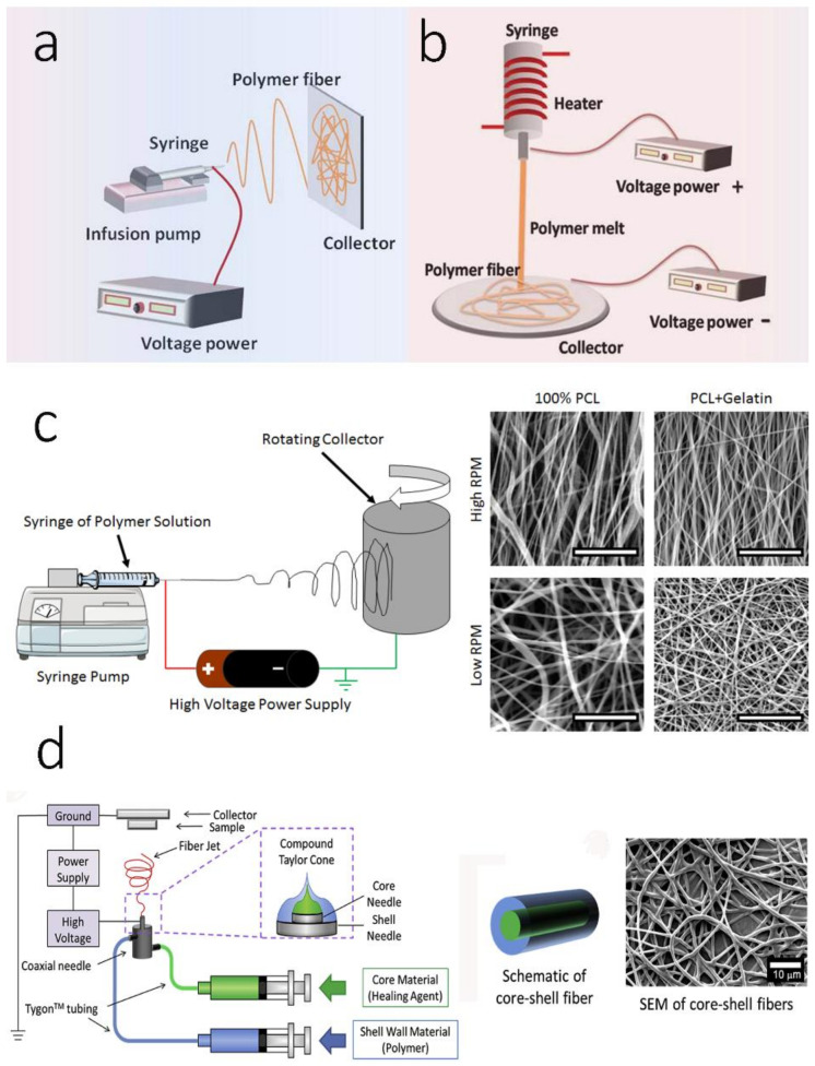
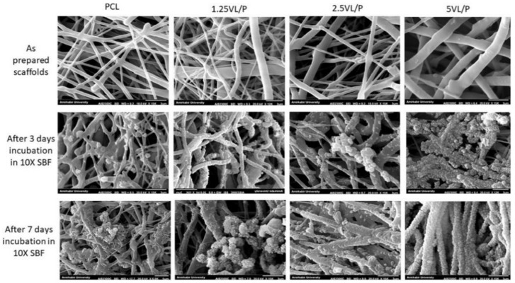
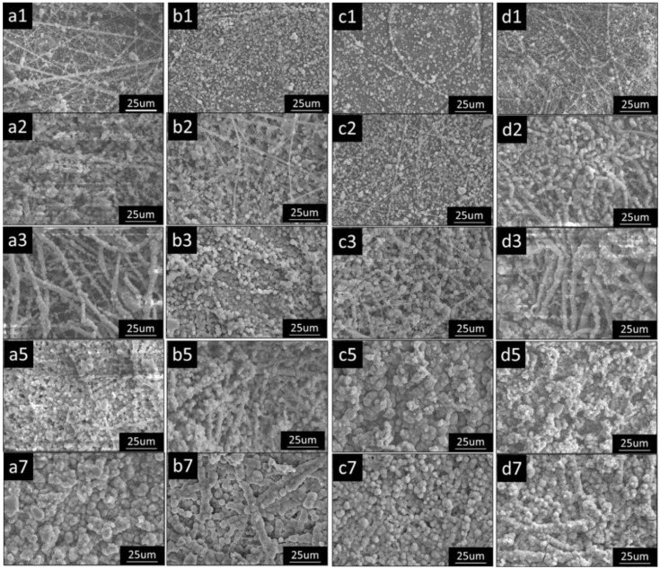
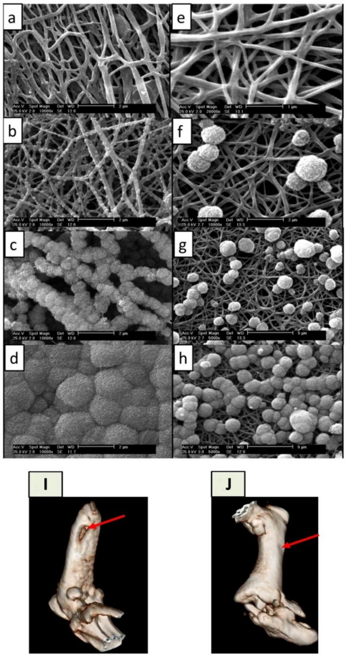
Increasing the demand for bone substitutes in the management of bone fractures, including osteoporotic fractures, makes bone tissue engineering (BTE) an ideal strategy for solving the constant shortage of bone grafts. Electrospun-based scaffolds have gained popularity in BTE because of their unique features, such as high porosity, a large surface-area-to-volume ratio, and their structural similarity to the native bone extracellular matrix (ECM). To imitate native bone mineralization through which bone minerals are deposited onto the bone matrix, a simple but robust post-treatment using a simulated body fluid (SBF) has been employed, thereby improving the osteogenic potential of these synthetic bone grafts. This study highlights recent electrospinning technologies that are helpful in creating more bone-like scaffolds, and addresses the progress of SBF development. Biomineralized electrospun bone scaffolds are also reviewed, based on the importance of bone mineralization in bone regeneration. This review summarizes the potential of SBF treatments for conferring the biphasic features of native bone ECM architectures onto electrospun-based bone scaffolds.

Keywords: bone mineralization; bone tissue engineering; electrospinning; simulated body fluid.


Conflict of interest statement

The author declares no conflict of interest.

Figures

Figure 1
Figure 1
Schematic image of bone anatomy. Reprinted with permission from Ref. [47]. Copyright 2017 MDPI. More details on “Copyright and Licensing” are available via the following link: https://www.mdpi.com/ethics#10, accessed on 15 April 2022.
Figure 2
Figure 2
Schematic images of different electrospun fabrication techniques: (a) monoaxial electrospinning; (b) melt electrospinning; (c) aligned electrospinning; (d) coaxial electrospinning. (a,b) reproduced with permission from Ref. [89]. Copyright 2017 Elsevier; (c) reproduced with permission from Ref. [90]. Copyright 2016 PLOS under a Creative Commons Attribution 4.0 International License; (d) reproduced with permission from Ref. [91]. Copyright 2016 Elsevier. More details on “Copyright and Licensing” are available via the following link: https://www.mdpi.com/ethics#10, accessed on 15 April 2022.
Figure 3
Figure 3
Scanning electron microscope images of as-prepared scaffolds and after 3 and 7 days incubation in 10× SBF. The formation of spherical apatite-like crystals increased significantly after adding nanohybrids to the scaffolds. For the legends, pure PCL and VD3·LDH/PCL electrospun scaffolds containing 1.25, 2.5, and 5 wt% of vitamin D3 are presented as PCL, 1.25VL/P, 2.5VL/P, and 5VL/P, respectively. Reprinted with permission from Ref. [139]. Copyright 2020 Elsevier. More details on “Copyright and Licensing” are available via the following link: https://www.mdpi.com/ethics#10, accessed on 15 April 2022.
Figure 4
Figure 4
Scanning electron microscope images of the gradual deposition of minerals onto PLLA/gelatin composite nanofibers over time in different concentrated SBF (2.5×) fortified with amino acids (2.5 mM) at 37 ± 0.2 °C. The numbers following the alphabets a–d indicate the soaking time (days). (a1a3,a5,a7) 2.5 SBF-blank; (b1b3,b5,b7) 2.5 SBF-Gly; (c1c3,c5,c7) 2.5 SBF-Arg; (d1d3,d5,d7) 2.5 SBF-Asp. The number shown in each panel of the figure represents the days of each SBF treatment. Magnification of 1000×. Reprinted with permission from Ref. [148]. Copyright 2015 Elsevier. More details on “Copyright and Licensing” are available via the following link: https://www.mdpi.com/ethics#10, accessed on 15 April 2022.
Figure 5
Figure 5
Scanning electron microscope (SEM) images of pristine carbon nanofibers (P-CNFs) and pre-treated carbon nanofibers (T-CNFs) incubated in a normal SBF. (a) T-CNFs-12 h, (b) T-CNFs-24 h, (c) T-CNFs-48 h, (d) T-CNFs-72 h, (e) P-CNFs-12 h, (f) P-CNFs-24, (g) P-CNFs-48 h, and (h) P-CNFs-72 h. (i,j) 3D computed tomography (CT) imaging of in vivo repair of a defective femur via mineralized carbon nanofibers (M-CNFs). Diagnostic 3D imaging (CT scan) of femur bone defects after 8 weeks of injury. The arrow shows the unrepaired defective site in the control group (i) and the bone defect repaired by normal tissue growth caused by the M-CNFs (j). Reprinted with permission from Ref. [156]. Copyright 2020 Nature publishing group. More details on “Copyright and Licensing” are available via the following link: https://www.mdpi.com/ethics#10, accessed on 15 April 2022.

Similar articles

Cited by

References

    1. Greenwald A.S., Boden S.D., Goldberg V.M., Khan Y., Laurencin C.T., Rosier R.N. Bone-Graft Substitutes: Facts, Fictions, and Applications. JBJS. 2001;83:S98–S103. doi: 10.2106/00004623-200100022-00007. - DOI - PubMed
    1. Rosen C.J. A Not-So-New Treatment for Old Bones. N. Engl. J. Med. 2018;379:2465–2466. doi: 10.1056/NEJMe1812434. - DOI - PubMed
    1. NIH Consensus Development Panel on Osteoporosis Prevention, Diagnosis, and Therapy Osteoporosis Prevention, Diagnosis, and Therapy. JAMA. 2001;285:785–795. doi: 10.1001/jama.285.6.785. - DOI - PubMed
    1. Föger-Samwald U., Dovjak P., Azizi-Semrad U., Kerschan-Schindl K., Pietschmann P. Osteoporosis: Pathophysiology and therapeutic options. EXCLI J. 2020;19:1017–1037. doi: 10.17179/excli2020-2591. - DOI - PMC - PubMed
    1. Tu K.N., Lie J.D., Wan C.K.V., Cameron M., Austel A.G., Nguyen J.K., Van K., Hyun D. Osteoporosis: A Review of Treatment Options. P T. 2018;43:92–104. - PMC - PubMed

LinkOut - more resources