

Segmentation of Plantar Foot Thermal Images Using Prior Information
- PMID: 35632244
- PMCID: PMC9146771
- DOI: 10.3390/s22103835
Segmentation of Plantar Foot Thermal Images Using Prior Information
Abstract
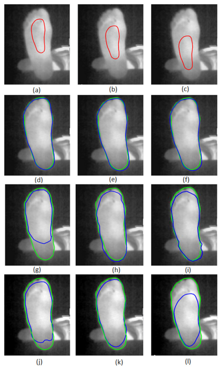
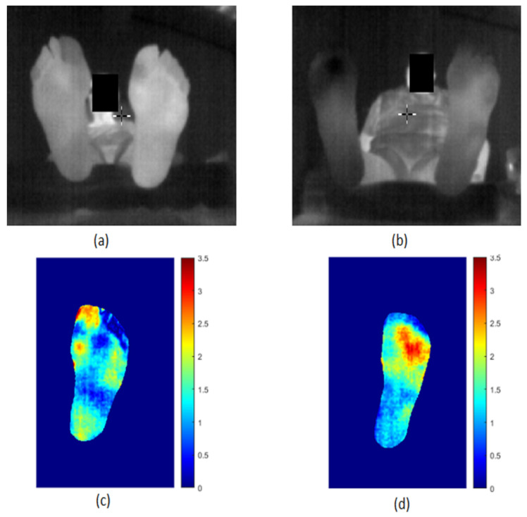
Diabetic foot (DF) complications are associated with temperature variations. The occurrence of DF ulceration could be reduced by using a contactless thermal camera. The aim of our study is to provide a decision support tool for the prevention of DF ulcers. Thus, the segmentation of the plantar foot in thermal images is a challenging step for a non-constraining acquisition protocol. This paper presents a new segmentation method for plantar foot thermal images. This method is designed to include five pieces of prior information regarding the aforementioned images. First, a new energy term is added to the snake of Kass et al. in order to force its curvature to match that of the prior shape, which has a known form. Second, we defined the initial contour as the downsized prior-shape contour, which is placed inside the plantar foot surface in a vertical orientation. This choice makes the snake avoid strong false boundaries present outside the plantar region when evolving. As a result, the snake produces a smooth contour that rapidly converges to the true boundaries of the foot. The proposed method is compared to two classical prior-shape snake methods, that of Ahmed et al. and that of Chen et al. A database of 50 plantar foot thermal images was processed. The results show that the proposed method outperforms the previous two methods with a root-mean-square error of 5.12 pixels and a dice similarity coefficient of 94%. The segmentation of the plantar foot regions in the thermal images helped us to assess the point-to-point temperature differences between the two feet in order to detect hyperthermia regions. The presence of such regions is the pre-sign of ulcers in the diabetic foot. Furthermore, our method was applied to hyperthermia detection to illustrate the promising potential of thermography in the case of the diabetic foot. Associated with a friendly acquisition protocol, the proposed segmentation method is the first step for a future mobile smartphone-based plantar foot thermal analysis for diabetic foot patients.
Keywords: active contours; diabetic foot; hyperthermia; infrared camera; plantar foot thermal images; prior-shape-based segmentation.
Conflict of interest statement
The authors declare no conflict of interest. The funders had no role in the design of the study; in the collection, analyses, or interpretation of data; in the writing of the manuscript, or in the decision to publish the results.
Figures


References
-
- Bouallal D., Bougrine A., Douzi H., Harba R., Canals R., Vilcahuaman L., Arbanil H. Segmentation of plantar foot thermal images: Application to diabetic foot diagnosis; Proceedings of the 2020 International Conference on Systems, Signals and Image Processing (IWSSIP); Niteroi, Brazil. 1–3 July 2020; pp. 116–121.
MeSH terms
Grants and funding
LinkOut - more resources
Full Text Sources
Medical
Research Materials
Miscellaneous
