Review
. 2022 May 10;10(5):751.
doi: 10.3390/vaccines10050751.

Vaccination against Bacterial Infections: Challenges, Progress, and New Approaches with a Focus on Intracellular Bacteria

Affiliations
Review

Vaccination against Bacterial Infections: Challenges, Progress, and New Approaches with a Focus on Intracellular Bacteria

Anke Osterloh. Vaccines (Basel). .

Abstract

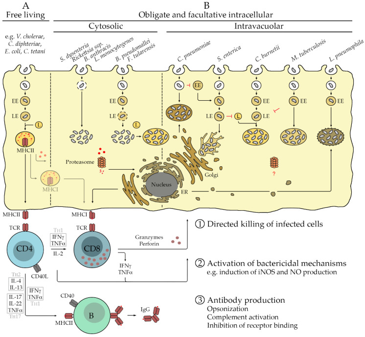
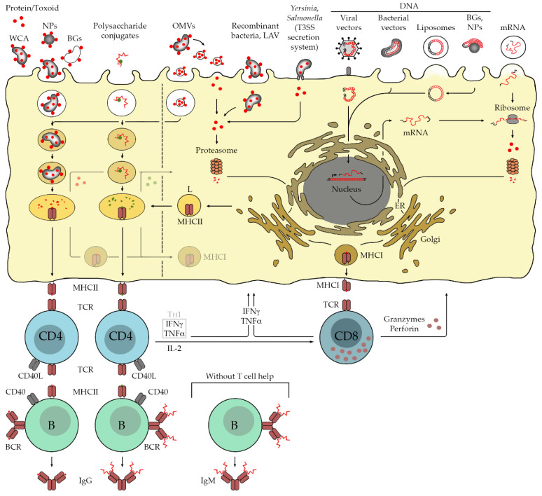
Many bacterial infections are major health problems worldwide, and treatment of many of these infectious diseases is becoming increasingly difficult due to the development of antibiotic resistance, which is a major threat. Prophylactic vaccines against these bacterial pathogens are urgently needed. This is also true for bacterial infections that are still neglected, even though they affect a large part of the world's population, especially under poor hygienic conditions. One example is typhus, a life-threatening disease also known as "war plague" caused by Rickettsia prowazekii, which could potentially come back in a war situation such as the one in Ukraine. However, vaccination against bacterial infections is a challenge. In general, bacteria are much more complex organisms than viruses and as such are more difficult targets. Unlike comparatively simple viruses, bacteria possess a variety of antigens whose immunogenic potential is often unknown, and it is unclear which antigen can elicit a protective and long-lasting immune response. Several vaccines against extracellular bacteria have been developed in the past and are still used successfully today, e.g., vaccines against tetanus, pertussis, and diphtheria. However, while induction of antibody production is usually sufficient for protection against extracellular bacteria, vaccination against intracellular bacteria is much more difficult because effective defense against these pathogens requires T cell-mediated responses, particularly the activation of cytotoxic CD8+ T cells. These responses are usually not efficiently elicited by immunization with non-living whole cell antigens or subunit vaccines, so that other antigen delivery strategies are required. This review provides an overview of existing antibacterial vaccines and novel approaches to vaccination with a focus on immunization against intracellular bacteria.

Keywords: antigens; extracellular and intracellular bacteria; immunity; vaccine.


Conflict of interest statement

The author declares no conflict of interest.

Figures

Figure 1
Figure 1
Extra- and intracellular bacteria and immune response. Free-living bacteria are taken up by phagocytes such as MØ and DCs as well as B cells that serve as professional antigen-presenting cells (APCs). Phagosomes develop into early endosomes (EE) and further to late endosomes (LE) that finally fuse with lysosomes (L). The activity of proteases and acidic environments of the Ls results in the degradation of the pathogen and its proteins, fragments of which are bound by MHCII molecules in the lysosomal membrane. MHCII/peptide complexes are presented on the cell surface to CD4+ T cells. The protective immune response is dominated by activated CD4+ T cells as well as B cells that produce antibodies against surface molecules of the pathogen. Depending on the cytokine environment provided by the APC, CD4+ T cells develop to T helper (TH) cells, either to TH2 cells producing IL-4 and IL-13, TH1 cells that secrete IFNγ and TNFα, or TH17 cells releasing IL-17, TNFα, and IL-22 that acts on non-immune cells. All TH cells also release IL-2, which promotes T cell proliferation and survival. Activated TH cells interact with activated B cells via the binding of CD40L to CD40 on the B cell surface, initiating the germinal center reaction where immunoglobulin class switch and affinity maturation occurs, so that high-affinity IgG instead of the initial IgM is produced. In addition, memory B cells develop. The cytokines that are produced by different TH cells promote the generation of certain IgG isotypes in this process. Antibodies can act against extracellular bacteria by opsonization for the uptake by phagocytes or direct destruction of the pathogen by complement activation. (A). Only a few bacteria replicate exclusively within target cells. These include members of the family of Rickettsiacea, Chlamydia (C.) pneumoniae, and Coxiella (C.) burnetii. Rickettsiae escape from the phagosome via the release of phospholipases that dissolve the phagosomal membrane and replicate free in the cytosol [6]. C. pneumoniae, instead, leaves the endocytic route and recruits Golgi-derived vesicles to form a unique compartment for replication that is associated with the microtubule organizing center (MTOC) [7]. Phagosomes containing C. burnetii, in turn, fuse with Ls to build a phagolysosomal-like vacuole for bacterial replication. Several other bacteria are facultative intracellular pathogens. Examples are B. anthracis, L. monocytogenes, B. pseudomallei, F. tularensis, S. enterica, M. tuberculosis, and L. pneumophila. Similar to rickettsiae, B. anthracis infects macrophages (MØ) and escapes from the phagosome to replicate free in the cytosol. L. monocytogenes, B. pseudomalleii, and F. tularensis deliberate from LEs and then also replicate free in the cytosol of infected cells. Cytosolic F. tularensis may also retranslocate into autolysosome-like vacuoles. In contrast, S. enterica inhibits fusion of LEs with Ls and replicates in LE-like compartments that are associated with the MTOC and form filaments [8]. M. tuberculosis inhibits maturation of EEs and replicates in LE-like vacuoles, while EEs containing L. pneumophila fuse with vesicles derived from the ER to form ribosome-coated compartments for bacterial replication [9]. (B). Efficient defense against intracellular pathogens usually requires the activation of cytotoxic CD8+ T cells that are capable of the direct killing of infected cells. CD8+ T cells are activated by antigenic peptides that derive from cytosolic proteins that are degraded by the proteasome. Peptides are transferred to the ER to be loaded onto MHCI molecules that are presented on the cell surface of all nucleated cells to be recognized by CD8+ T cells. Bacterial antigens that are recognized by CD8+ T cells may derive predominantly from secreted proteins or surface proteins that are accessible for proteasomal degradation in the cytosol ①. Initial activation of CD8+ T cells and defense against intracellular bacteria further require the activation of CD4+ T cells, predominantly of the TH1 type. These cells support CD8+ T cell responses. In addition, CD4+ TH1 cells (as well as TH17 cells and IFNγ-releasing CD8+ T cells) induce bactericidal mechanisms such as the induction of nitric oxide synthase (iNOS) and NO production in infected cells via the release of IFNγ and TNFα. In this way, CD4+ TH1 cells contribute to bacterial elimination ②. Antibodies produced by B cells may play a minor role in the defense against primary infection with intracellular bacteria but can contribute to protection in secondary infection. In addition to the aforementioned mechanisms, antibodies can here participate in defense by the inhibition of the binding of the bacteria to receptors that mediate bacterial uptake into target cells ③. For those bacteria that replicate within cellular compartments and thus are hidden from the cytosol and the proteasome, the activation of CD8+ T cell responses during the infection may not be efficient.
Figure 2
Figure 2
Applied and experimental bacterial vaccines. Today, established bacterial vaccines that are in use are the immunizations with WCA, recombinant proteins including toxoids, polysaccharide/protein conjugates, live attenuated vaccines (LAV), and, since the last few years, also bacterial outer membrane vesicles (OMVs). Vaccination with WCA, bacterial ghosts (BGs), and recombinant proteins/toxoids predominantly results in the processing of protein components for the presentation via MHCII and the activation predominantly of CD4+ T cells. In addition, antigen-specific activated B cells produce high-affinity IgG antibodies with the help of CD4+ T cells, and a memory response is induced. In the case of polysaccharide/protein conjugates, the carrier protein serves as the protein component that can be recognized by CD4+ T cells. This enables B cells to produce high-affinity IgG antibodies against the polysaccharide instead of the production of low-affinity IgM without T cell help. Immunization against intracellular bacterial pathogens requires the activation of cytotoxic CD8+ T cells, which is usually not efficiently achieved with these methods. Antigens recognized by CD8+ T cells predominantly derive from cytosolic proteins that are degraded by the proteasome and further processed for the presentation via MHCI. A major difficulty of efficient vaccination against intracellular bacterial pathogens lies in the introduction of immunogens into the cytosol of host cells. This can be achieved by the use of OMVs, LAV, viral vectors, bacterial vectors, immunization with DNA or mRNA, and the use of the T3SS translocation system of bacteria such as Salmonella. Immunization with OMVs and LAV results in both antigen presentation by MHCII and MHCI molecules for the activation of CD4+ and CD8+ T cells, whereby the mechanisms of MHCI presentation in the case of OMV immunization are not well understood and may be a result of cross presentation, either by the release of proteins from the lysosome into the cytosol or by fusion of lysosomes with MHCI-containing vesicles. LAV may release proteins into the cytosol. In addition, surface proteins may be accessible to the proteasome for processing via the MHCI presentation pathway. The most frequently used viral vectors for vaccination are adenoviruses and modified vaccinia virus Ankara (MVA). Adenoviruses translocate their double-stranded (ds) DNA genome into the nucleus of non-dividing cells for replication. Viral mRNA transcription products are exported into the cytosol of the infected cells where ribosomal translation occurs. In contrast, MVA, which also carries a dsDNA genome, has a unique replication cycle that is restricted to the cytosol. In both cases, proteins are expressed in the cytosol of infected cells, which has also been shown for bacterial vectors that carry plasmid DNA with eukaryotic expression cassettes for the expression of immunogens. Cytosolic protein expression is also achieved by the direct transfection of target cells with either DNA or mRNA. While mRNA is transferred directly into the cytosol for protein translation, DNA has to enter the nucleus of the target cell for transcription, which is usually more efficiently achieved with viral vectors. Finally, a rather experimental approach to the introduction of antigens into the cytosol of target cells is the use of recombinant attenuated bacteria such as Salmonella that possess a T3SS translocation system. This system allows active and direct injection of proteins into the cytosol of target cells. In contrast to the use of WCA and LAV, all other methods generally require knowledge of the immunogenic determinants of the pathogen to prepare recombinant vaccines.

Similar articles

Cited by

References

    1. Dramsi S., Cossart P. Intracellular pathogens and the actin cytoskeleton. Annu. Rev. Cell Dev. Biol. 1998;14:137–166. doi: 10.1146/annurev.cellbio.14.1.137. - DOI - PubMed
    1. Mitchell G., Chen C., Portnoy D.A. Strategies Used by Bacteria to Grow in Macrophages. Microbiol. Spectr. 2016;4:701–725. doi: 10.1128/microbiolspec.MCHD-0012-2015. - DOI - PMC - PubMed
    1. Chong A., Wehrly T.D., Nair V., Fischer E.R., Barker J.R., Klose K.E., Celli J. The Early Phagosomal Stage of Francisella tularensis Determines Optimal Phagosomal Escape and Francisella Pathogenicity Island Protein Expression. Infect. Immun. 2008;76:5488–5499. doi: 10.1128/IAI.00682-08. - DOI - PMC - PubMed
    1. Baca O.G., Li Y.-P., Kumar H. Survival of the Q fever agent Coxiella burnetii in the phagolysosome. Trends Microbiol. 1994;2:476–480. doi: 10.1016/0966-842X(94)90651-3. - DOI - PubMed
    1. Steele-Mortimer O., Meresse S., Gorvel J.-P., Toh B.-H., Finlay B.B. Biogenesis of Salmonella typhimurium-containing vacuoles in epithelial cells involves interactions with the early endocytic pathway. Cell. Microbiol. 1999;1:33–49. doi: 10.1046/j.1462-5822.1999.00003.x. - DOI - PubMed