Review
. 2022 May 12;10(5):770.
doi: 10.3390/vaccines10050770.

Persistent Enterovirus Infection: Little Deletions, Long Infections

Affiliations
Review

Persistent Enterovirus Infection: Little Deletions, Long Infections

Nora M Chapman. Vaccines (Basel). .

Abstract

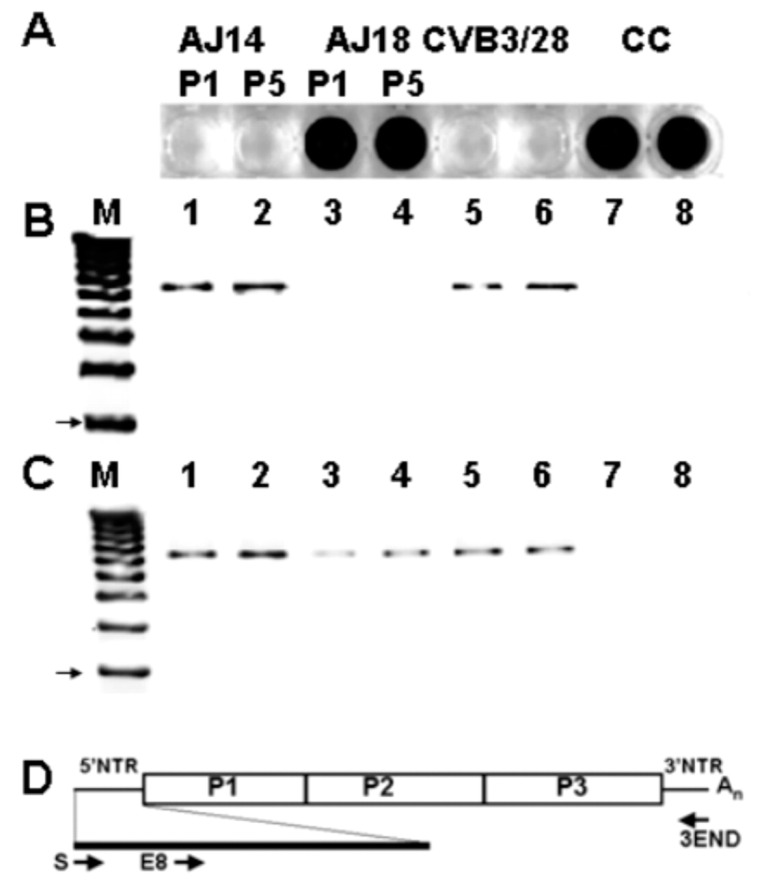
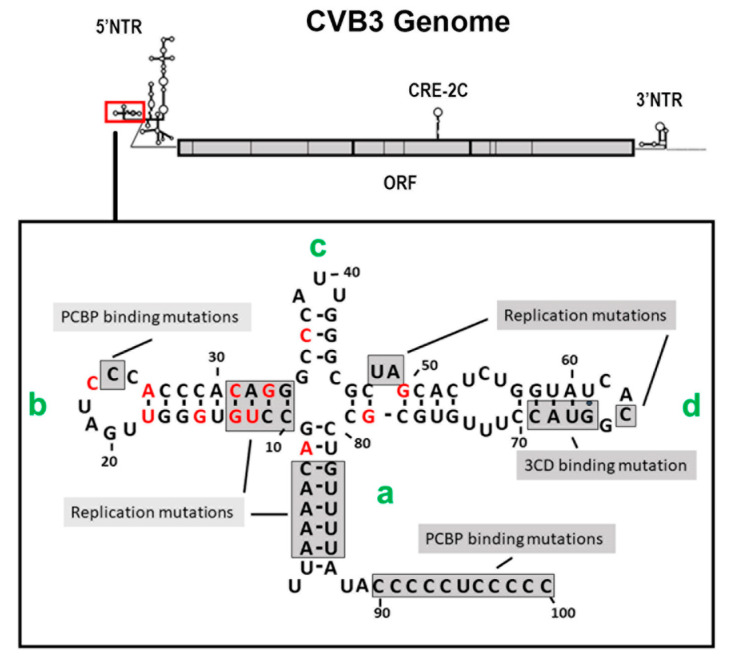
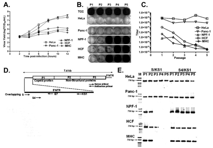
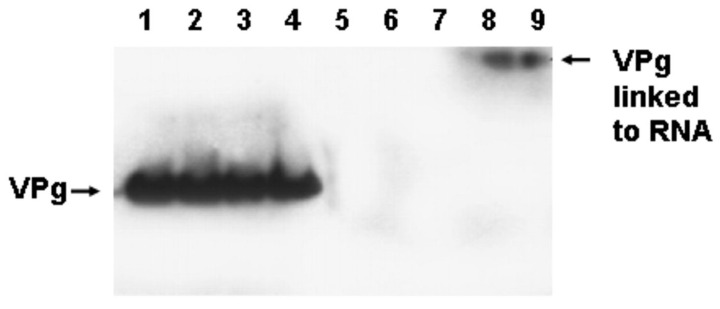
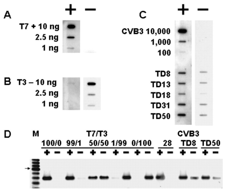
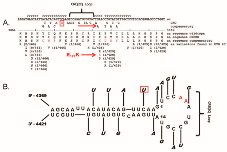
Enteroviruses have now been shown to persist in cell cultures and in vivo by a novel mechanism involving the deletion of varying amounts of the 5' terminal genomic region termed domain I (also known as the cloverleaf). Molecular clones of coxsackievirus B3 (CVB3) genomes with 5' terminal deletions (TD) of varying length allow the study of these mutant populations, which are able to replicate in the complete absence of wildtype virus genomes. The study of TD enteroviruses has revealed numerous significant differences from canonical enteroviral biology. The deletions appear and become the dominant population when an enterovirus replicates in quiescent cell populations, but can also occur if one of the cis-acting replication elements of the genome (CRE-2C) is artificially mutated in the element's stem and loop structures. This review discusses how the TD genomes arise, how they interact with the host, and their effects on host biology.

Keywords: cis-acting replication element; coxsackievirus B; enterovirus; negative-strand initiation; persistent infection; positive-strand initiation; terminal deletion.


Conflict of interest statement

The author declares no conflict of interest.

Figures

Figure 1
Figure 1
Infection of HeLa cells with homogenate of CVB3-infected AJ heart at day 18 does not induce cytopathic effect, but does replicate CVB3 RNA. (A) HeLa cultures inoculated with homogenates of hearts at days 14 p.i. (AJ14) and 18 p.i. (AJ18) passed once (P1) and five times (P5) in HeLa inoculated with parental virus (CVB3/28) or uninfected cell culture (CC). Cells were stained with crystal violet. (B,C) Total RNA purified from HeLa-passaged AJ14, AJ18, CVB3/28-inoculated HeLa cells, and uninfected cells was used for RT-PCR with the primer 3END and with primers S (B) or E8 (C). The arrow indicates a 3 kb band. Lane M, 1 kb DNA ladder; lanes 1 and 2, AJ14 P1 and P5, respectively; lanes 3 and 4, AJ18 P1 and P5, respectively; lanes 5 and 6, CVB3/28; lanes 7 and 8, uninfected cell culture. (D) Relative positions in the CVB3 genome of primers used. Reproduced with permission from [50] 2005, American Society for Microbiology.
Figure 2
Figure 2
Cloverleaf of CVB3. Green letters (a, b, c, and d) refer to stem and stem-loops. Red letters indicate the first nucleotide of CVB-TDs generated in cell culture or in vivo. Shaded regions indicate sites of mutations in enterovirus genomes.
Figure 3
Figure 3
CVB3 replicates, but does not induce, CPE in the indicator HeLa cell monolayers following sequential infection of replication-restricted primary cells. (A) Single-step growth curves of CVB3/28 in primary cell cultures NPF-1, MHC, and HCF, and in immortal cell lines HeLa and Panc-1 (inoculated with an MOI of 20 TCID50 per cell). Error bars show standard deviations. (B) Supernatants of CVB3/28 passaged in primary cell cultures lose the ability to cause CPE in HeLa cell monolayers. P1 to P5, HeLa cells infected with supernatants of passages 1 to 5; TCC, uninfected HeLa cell control. Cell cultures were fixed in acetic acid/acetone and then stained with crystal violet. Titers at each passage were determined either by CPE on HeLa cell monolayers or by real-time quantitative RT-PCR. (C) Titers from serial passages shown in panel B are plotted. (D) Relative positions in the CVB3 genome of primers used in this study. (E) CVB3/28 RNAs in primary cell cultures detected by RT-PCR. Lane M, Hi-Lo DNA marker (Minnesota Molecular, Inc., Minneapolis, MN); lanes P1 to P5, passages 1 to 5. Arrows indicate 750 bp bands. Reprinted with permission from [61], 2008, American Society for Microbiology.
Figure 4
Figure 4
CVB3/TD viruses have VPg covalently attached to genomic RNA. Viral RNA was purified from stocks of the CVB3/28, TD8, and TD50 viruses (105 TCID50 units) and analyzed by Western blotting. Blots were probed with an antibody to PV1 VPg (N10). Lane 1 (PV1 Sabin), lane 2, 5 (CVB3/28), lane 3, 6, 8 (CVB3/TD8), lane 4, 7, 9 (CVB3/TD50). Lanes 1–4, RNA was treated with RNase A/T1; lanes 5–7, RNA was treated with RNase A/T1 and proteinase K; lanes 8 and 9, RNA was untreated. Adapted with permission from [50] 2005, American Society for Microbiology.
Figure 5
Figure 5
Positive and negative strands in wildtype and TD virions. (AC) Slot blots of control and virion RNA detected with labeled strand-specific oligonucleotides. (D) Purified strands of control mixtures of strands or virion RNA amplified with RT-PCR and agarose electrophoresed. Reprinted with permission from [50], 2005, American Society for Microbiology.
Figure 6
Figure 6
Compensatory mutations are unlikely in the stem of the CRE-2C. (A) Sequence analysis using NCBI was used to explore the possibility of compensatory mutations in the CRE-2C of CVB3. Potential amino acid variations resulting from compensatory mutations to restore the CRE-2C RNA structure are noted. All variations in the CRE-2C-encoding sequence that would affect the amino acid sequence of the 2C protein are noted. Only 1 amino acid variation identical to those resulting from compensatory mutations was reported to exist in 429 EV-B 2C aa sequences examined (box and arrows). (B) Location of this compensatory mutation in the CRE-2C secondary structure (box). Reprinted with permission [77], 2016, Elsevier.

References

    1. Racaniello V.R. Picornaviridae: The Viruses and their Replication. In: Knipe D.M., Howley P.M., Cohen J.I., Griffin D.E., Lamb R.A., Martin M.A., Racaniello V.R., Roizman B., editors. Fields Virology. 6th ed. Lippincott Williams & Wilkins; Philadelphia, PA, USA: 2013. pp. 453–489.
    1. Pallansch M.A., Oberste M.S., Whitton J.L. Enteroviruses: Polioviruses, Coxsackieviruses, Echoviruses, and Newer Enteroviruses. In: Knipe D.M., Howley P.M., Cohen J.I., Griffin D.E., Lamb R.A., Martin M.A., Racaniello V.R., Roizman B., editors. Fields Virology. 6th ed. Lippincott Williams & Wilkins; Philadelphia, PA, USA: 2001. pp. 490–530.
    1. Kim K.-S., Chapman N., Tracy S. The group B coxsackieviruses and myocarditis. Rev. Med. Virol. 2001;11:355–368. doi: 10.1002/rmv.326. - DOI - PubMed
    1. Lloyd R.E., Tamhankar M., Lernmark A. Enteroviruses and Type 1 Diabetes: Multiple Mechanisms and Factors? Annu. Rev. Med. 2022;73:483–499. doi: 10.1146/annurev-med-042320-015952. - DOI - PMC - PubMed
    1. Heim A., Canu A., Kirschner P., Simon T., Mall G., Hofschneider P.H., Kandolf R. Synergistic Interaction of Interferon- and Interferon- in Coxsackievirus B3-Infected Carrier Cultures of Human Myocardial Fibroblasts. J. Infect. Dis. 1992;166:958–965. doi: 10.1093/infdis/166.5.985. - DOI - PubMed

LinkOut - more resources