Review
. 2022 Apr 28;14(5):926.
doi: 10.3390/v14050926.

Multimodal Functionalities of HIV-1 Integrase

Affiliations
Review

Multimodal Functionalities of HIV-1 Integrase

Alan N Engelman et al. Viruses. .

Abstract

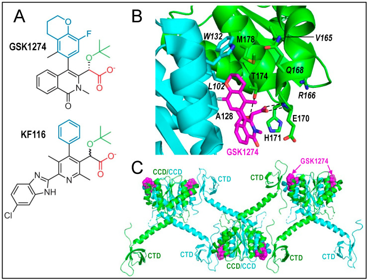
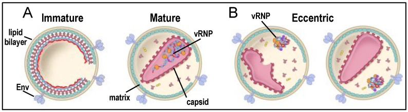
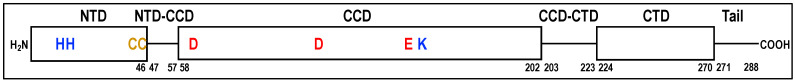
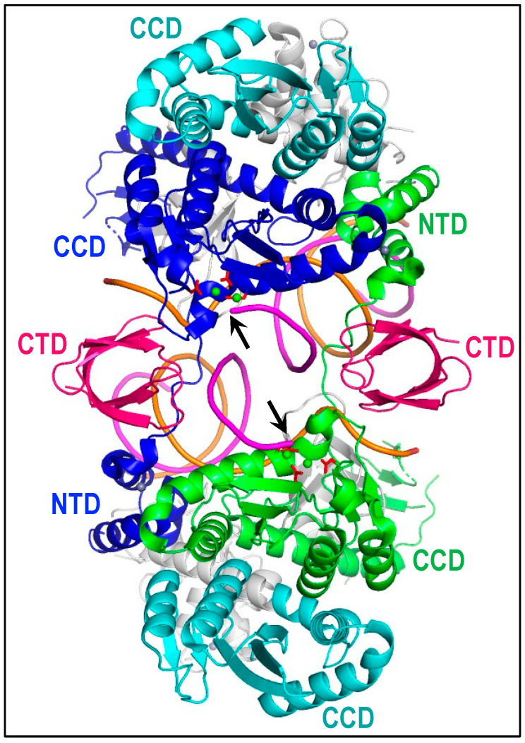
Integrase is the retroviral protein responsible for integrating reverse transcripts into cellular genomes. Co-packaged with viral RNA and reverse transcriptase into capsid-encased viral cores, human immunodeficiency virus 1 (HIV-1) integrase has long been implicated in reverse transcription and virion maturation. However, the underlying mechanisms of integrase in these non-catalytic-related viral replication steps have remained elusive. Recent results have shown that integrase binds genomic RNA in virions, and that mutational or pharmacological disruption of integrase-RNA binding yields eccentric virion particles with ribonucleoprotein complexes situated outside of the capsid shell. Such viruses are defective for reverse transcription due to preferential loss of integrase and viral RNA from infected target cells. Parallel research has revealed defective integrase-RNA binding and eccentric particle formation as common features of class II integrase mutant viruses, a phenotypic grouping of viruses that display defects at steps beyond integration. In light of these new findings, we propose three new subclasses of class II mutant viruses (a, b, and c), all of which are defective for integrase-RNA binding and particle morphogenesis, but differ based on distinct underlying mechanisms exhibited by the associated integrase mutant proteins. We also assess how these findings inform the role of integrase in HIV-1 particle maturation.

Keywords: HIV; aberrant integrase multimerization; allosteric integrase inhibitor; integrase; integrase-RNA binding; virus maturation; virus morphogenesis.


Conflict of interest statement

A.N.E. has in the past received fees from ViiV Healthcare, Co. M.K. declares no conflict of interest.

Figures

Figure 6
Figure 6
Representative ALLINI chemotypes and IN binding modes. (A) Structures of quinoline GSK1274 [222] and pyridine KF116 [223] compounds. See text for color descriptors. (B) X-ray crystal structure of GSK1274 bound to the IN CCD dimer (PDB accession code 4OJR [222]); the IN protein harbored the solubility-enhancing F185K substitution [74,232]. CCD dimerization interface residues in close contact to GSK1264 are shown. Nearby Val165 and Arg166 are additionally highlighted. Amino acid residues that when mutated yield class IIb (Val165, Gln168) or class IIc (Leu102, Trp132, Arg166) IN mutant viruses are labeled in italics. Atoms are colored as follows: red, oxygen; blue, nitrogen; mustard, sulfur; and light blue, fluorine. Dashed lines, hydrogen bond interactions. (C) Structure depicting ALLINI-induced IN hyper-multimerization (PDB accession code 5HOT [230]). The full-length IN construct harbored Y15A and F185H amino acid substitutions. The IN NTD was not resolved in the electron density map; other domains are indicated. GSK1264 is colored magenta in panels B and C.
Figure 1
Figure 1
HIV-1 virion particles. (A) Schematic of immature and mature virions. Note the vRNP encased in the capsid shell in mature particles. (B) Eccentric virions with deformed (left) or closed (right) capsid. Env, envelope glycoproteins. See main text for additional descriptions. Adapted from Ref. [7].
Figure 2
Figure 2
HIV-1 IN domains and phylogenetically conserved amino acid residues. Upper labels denote domains, interdomain linkers, and C-terminal tail region. Active site DDE residues are colored red, nitrogen-containing side chains blue, and cysteines are colored dark yellow. CCD residue Lys159, which is conserved among retroviral but not retrotransposon INs, interacts with the vDNA end [75,76]. Numbers, based on Refs. [77,78], denote domain and linker boundaries.
Figure 3
Figure 3
HIV-1 intasome structure (protein database (PDB) accession code 6PUT). Cyan-blue (top) and green-cyan (bottom) IN dimers interact via the CCD-CCD dimerization interface. The internal blue-green IN dimer is bound to vDNA, which is colored orange and magenta to highlight non-transferred and transferred DNA strands, respectively. The 3′ ends of the magenta transferred strands, which are indicated by arrows, are engaged by inner IN dimer DDE active sites (red sticks in association with two calcium ions, which are shown as green spheres). NTD-associated grey spheres are zinc. Synaptic CTDs are in hotpink to indicate their origin from peripheral IN tetramers, which were otherwise omitted from the atomic model. Gray is used to deemphasize secondary structural elements that do not directly contribute to the CIC.
Figure 4
Figure 4
Cartoon representation of the CTD and six downstream residues (amino acids 271–276) of the IN tail region (PDB accession code 5HOT). The side chains of IN residues Lys264, Lys266, Arg269, and Lys273, which are implicated in vRNA binding, are shown as sticks.
Figure 5
Figure 5
Class I and class II IN mutations and IN oligomerization interface proximity. (A) The blue CCD from the HIV-1 intasome in Figure 3 was resected and positioned to highlight active site proximal amino acid residues (in yellow space fill) that when changed yield the class I IN mutant viral phenotype. DDE active site residues are shown as red sticks. (B) The panel A image was rotated clockwise ~45° around the y-axis to highlight the cyan CCD-blue CCD dimerization interface. Blue CCD and green NTD amino acid residues that when mutated yield the class IIc IN mutant viral phenotype (see Table 1, below) are shown in space fill [51,56,122,124,148,149,150]; His12, His16, and Cys40 are collectively labeled HHCC. The panel B image highlights oligomerization interface proximity of residues that when changed yield class II IN mutant viruses. For clarity, the DNA duplex was omitted from panel B.

References

    1. Giron M.L., Colas S., Wybier J., Rozain F., Emanoil-Ravier R. Expression and maturation of human foamy virus Gag precursor polypeptides. J. Virol. 1997;71:1635–1639. doi: 10.1128/jvi.71.2.1635-1639.1997. - DOI - PMC - PubMed
    1. Effantin G., Estrozi L.F., Aschman N., Renesto P., Stanke N., Lindemann D., Schoehn G., Weissenhorn W. Cryo-electron microscopy structure of the native prototype foamy virus glycoprotein and virus architecture. PLoS Pathog. 2016;12:e1005721. doi: 10.1371/journal.ppat.1005721. - DOI - PMC - PubMed
    1. Qu K., Ke Z., Zila V., Anders-Össwein M., Glass B., Mücksch F., Müller R., Schultz C., Müller B., Kräusslich H.G., et al. Maturation of the matrix and viral membrane of HIV-1. Science. 2021;373:700–704. doi: 10.1126/science.abe6821. - DOI - PMC - PubMed
    1. Ganser B.K., Li S., Klishko V.Y., Finch J.T., Sundquist W.I. Assembly and analysis of conical models for the HIV-1 core. Science. 1999;283:80–83. doi: 10.1126/science.283.5398.80. - DOI - PubMed
    1. Zhao G., Perilla J.R., Yufenyuy E.L., Meng X., Chen B., Ning J., Ahn J., Gronenborn A.M., Schulten K., Aiken C., et al. Mature HIV-1 capsid structure by cryo-electron microscopy and all-atom molecular dynamics. Nature. 2013;497:643–646. doi: 10.1038/nature12162. - DOI - PMC - PubMed

Publication types

Substances

LinkOut - more resources