Case Reports
. 2022 May 8;14(5):1000.
doi: 10.3390/v14051000.

A Novel In Vivo Model of Laryngeal Papillomavirus-Associated Disease Using Mus musculus Papillomavirus

Affiliations
Case Reports

A Novel In Vivo Model of Laryngeal Papillomavirus-Associated Disease Using Mus musculus Papillomavirus

Renee E King et al. Viruses. .

Abstract

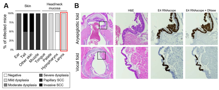
Recurrent respiratory papillomatosis (RRP), caused by laryngeal infection with low-risk human papillomaviruses, has devastating effects on vocal communication and quality of life. Factors in RRP onset, other than viral presence in the airway, are poorly understood. RRP research has been stalled by limited preclinical models. The only known papillomavirus able to infect laboratory mice, Mus musculus papillomavirus (MmuPV1), induces disease in a variety of tissues. We hypothesized that MmuPV1 could infect the larynx as a foundation for a preclinical model of RRP. We further hypothesized that epithelial injury would enhance the ability of MmuPV1 to cause laryngeal disease, because injury is a potential factor in RRP and promotes MmuPV1 infection in other tissues. In this report, we infected larynges of NOD scid gamma mice with MmuPV1 with and without vocal fold abrasion and measured infection and disease pathogenesis over 12 weeks. Laryngeal disease incidence and severity increased earlier in mice that underwent injury in addition to infection. However, laryngeal disease emerged in all infected mice by week 12, with or without injury. Secondary laryngeal infections and disease arose in nude mice after MmuPV1 skin infections, confirming that experimentally induced injury is dispensable for laryngeal MmuPV1 infection and disease in immunocompromised mice. Unlike RRP, lesions were relatively flat dysplasias and they could progress to cancer. Similar to RRP, MmuPV1 transcript was detected in all laryngeal disease and in clinically normal larynges. MmuPV1 capsid protein was largely absent from the larynx, but productive infection arose in a case of squamous metaplasia at the level of the cricoid cartilage. Similar to RRP, disease spread beyond the larynx to the trachea and bronchi. This first report of laryngeal MmuPV1 infection provides a foundation for a preclinical model of RRP.

Keywords: MmuPV1; RRP; laryngoscopy; larynx; mouse; papillomavirus; recurrent respiratory papillomatosis; vocal folds.


Conflict of interest statement

Sales of the mouse anesthesia nose cone are licensed through the Wisconsin Alumni Research Foundation. The inventors (including A.B. and P.F.L.) will receive a portion of the proceeds. R.E.K., J.R., E.T.W-S., R.H. and S.L.T. declare no conflicts of interest. The funders had no role in the design of the study; in the collection, analyses, or interpretation of data; in the writing of the manuscript; or in the decision to publish the results.

Figures

Figure 1
Figure 1
Study design.
Figure 2
Figure 2
Murine endoscopic laryngeal procedures. (A) Endoscopic images of pharynx and larynx with anatomic orientation and relevant tissues labeled, progressing from the back of the oral cavity (cranial) to the larynx (caudal). (B) Custom stainless steel micro brush for vocal fold abrasion with 0.5 mm diameter. (C) Gastight 10 uL syringe, 26-gauge needle, and 26-gauge PTFE tubing within the assembled operating sheath and endoscope. (D) Endoscopic image of murine vocal fold abrasion. Brush bristles are apparent between spiral wire. (E) Endoscopic image of murine vocal fold inoculation. Drops of inoculate are visible within tubing. Clear tubing appears white due to image magnification and reflected light.
Figure 3
Figure 3
Gross and microscopic murine vocal fold lesions post MmuPV1 infection. H&E-stained histologic and endoscopic images of vocal folds from the same animal. (A) Week 12 post saline mock infection. (B) Week 8 post MmuPV1 infection. Arrows indicate grossly visible, smooth vocal fold lesion. (C) Week 12 post vocal fold abrasion and MmuPV1 infection. Arrows indicate grossly visible, smooth, pale vocal fold lesion. (D) Week 4 post MmuPV1 infection. Arrow indicates lesion on histology. Lesion was not visible on endoscopy.
Figure 4
Figure 4
MmuPV1 induced severe laryngeal disease. Infected groups only, n = 15–20 mice per timepoint. (A) Key for panels (B)–(E). (B) Disease incidence in larynx for each animal. (C) Highest disease severity in larynx for each animal. (D) Disease incidence in larynx vs. other head and neck tissues. Fisher’s exact tests of differences in disease incidence among head/neck tissues at each timepoint: week 1, p = 0.2976; week 2, p < 0.0001; week 4, p = 0.0001; week 8, p < 0.0001; week 12, p < 0.0001. * Significant post hoc Bonferroni-adjusted Fisher’s exact pairwise tests between the larynx and other tissues: larynx vs. trachea week 2, p = 0.0080; week 4, p = 0.0079; week 8, p < 0.0001; week 12, p < 0.0001. All other pairwise tests vs. larynx: p > 0.05/4 = 0.0125. (E) Disease severity in larynx vs. other head and neck tissues. Severity was coded ordinally for analysis: negative = 0, mild dysplasia = 1, moderate dysplasia = 2, severe dysplasia = 3, invasive cancer = 4. Kruskal–Wallis tests of severity among tissues at each timepoint: week 1, p = 0.2480; week 2, p < 0.0001; week 4, p = 0.0001; week 8, p < 0.0001; week 12, p < 0.0001. * Significant post hoc DSCF test between larynx and other tissues: week 2 larynx vs. trachea p = 0.0322; week 4 larynx vs. hypopharynx p = 0.0121, larynx vs. tongue p = 0.0340; week 8 larynx vs. trachea p = 0.0003, larynx vs. hypopharynx p = 0.0140, larynx vs. tongue p = 0.0006, larynx vs. palate p = 0.0020; week 12 larynx vs. trachea p < 0.0001, larynx vs. hypopharynx p < 0.0001, larynx vs. tongue p < 0.0001, larynx vs. palate p < 0.0001. All other pairwise tests vs. larynx: adjusted p > 0.05.
Figure 5
Figure 5
Injury enhanced, but was not necessary, for MmuPV1-induced laryngeal disease in NSG mice. Infected groups week 1 and later only, n = 7–10 mice per group per timepoint. (A) Laryngeal disease incidence after laryngeal MmuPV1 infection vs. vocal fold abrasion and infection. * Significant Fisher’s exact test of group difference in dysplasia incidence in the larynx: week 4, p = 0.0198. All other p > 0.05. (B) Laryngeal disease severity after laryngeal MmuPV1 infection vs. vocal fold abrasion and infection. Severity was coded ordinally: negative = 0, mild dysplasia = 1, moderate dysplasia = 2, severe dysplasia = 3, invasive cancer = 4. * One-tailed Wilcoxon rank-sum tests of severity in Abrasion + MmuPV1 vs. MmuPV1 at each timepoint: week 4, p = 0.0083; week 8, p = 0.0313. All other p > 0.05.
Figure 6
Figure 6
MmuPV1-induced vocal fold dysplasia was positive for E4 transcript and negative for L1 capsid protein, regardless of severity. (A) Low magnification of H&E-stained coronal section of murine larynx. Box indicates vocal folds. (B) Serial sections of mild, moderate, and severe vocal fold dysplasia stained with H&E, MmuPV1 E4 RNAscope ISH, and MmuPV1 L1/K14 IF. 40×.
Figure 7
Figure 7
Squamous metaplasia and severe dysplasia at the level of the cricoid cartilage was positive for MmuPV1 transcripts and L1 capsid protein. (A) Low magnification of H&E-stained coronal section of murine larynx and 40× magnifications of area indicated in box and serial slides stained with H&E, MmuPV1 L1/K14 IF, and MmuPV1 L1/K14 IF with no L1 primary antibody. (B) Same stains as in (A), 10 slides (20 sections) more posterior within the larynx. (C) Sections serial to (A), stained with MmuPV1 E4 RNAscope ISH, E4 RNAscope with DNase treatment, E4 RNAscope with DNase and RNase treatment, and negative control probe RNAscope ISH.
Figure 8
Figure 8
Subclinical MmuPV1 infections in larynx, trachea, and hypopharynx day 1 and 3 after infection. Low magnification of H&E-stained coronal sections of murine larynx and 40× magnifications of areas indicated in boxes with serial sections stained with H&E, MmuPV1 E4 RNAscope ISH, E4 RNAscope with DNase treatment, E4 RNAscope with DNase and RNase treatment, and negative control probe RNAscope ISH.
Figure 9
Figure 9
MmuPV1 capsid was produced in the infected palate, but not in the nasopharynx. (A) Low magnification of an H&E-stained sagittal section of a murine palate. Boxes indicate areas magnified in (B,C). (B) 40× magnification of solid box in (A). Serial sections of mild sinonasal dysplasia stained with H&E, MmuPV1 E4 ISH, and MmuPV1 L1/K14 IF. (C) 40× magnification of dashed box in (A). Serial sections of mild palate dysplasia stained with H&E, MmuPV1 E4 ISH, and MmuPV1 L1/K14 IF.
Figure 10
Figure 10
MmuPV1 infection spread to the distal trachea and bronchi, but not to the lung. (A) Gel electrophoresis of the PCR results for MmuPV1 E2 and Mus musculus p53 in distal tracheal tissue DNA at week 12 post procedure. (B,C) Low magnification of an H&E-stained section of murine lungs and 40× magnification of areas indicated with arrows, with serial sections stained with H&E, MmuPV1 E4 RNAscope ISH, and E4 RNAscope with DNase treatment. (B) Bronchus positive for E4. (C) Lung with questionable E4 signal. (D) Gel electrophoresis of the PCR results for MmuPV1 E2 and Mus musculus p53 in DNA isolated from 2 slides/4 sections of 5 um coronal lung.
Figure 11
Figure 11
Secondary laryngeal MmuPV1 infections and dysplasias in nude mice arose after skin infection with laryngeal lavage. (A) Disease severity in nude mice infected with laryngeal lavage. SCC: squamous cell carcinoma. (B) Low magnification of H&E-stained sections of murine larynges and 40× magnification of areas indicated with boxes, with serial sections stained with H&E, MmuPV1 E4 RNAscope ISH, and E4 RNAscope with DNase treatment.

References

    1. Larson D.A., Derkay C.S. Epidemiology of Recurrent Respiratory Papillomatosis. APMIS. 2010;118:450–454. doi: 10.1111/j.1600-0463.2010.02619.x. - DOI - PubMed
    1. Fortes H.R., von Ranke F.M., Escuissato D.L., Araujo Neto C.A., Zanetti G., Hochhegger B., Souza C.A., Marchiori E. Recurrent Respiratory Papillomatosis: A State-of-the-Art Review. Respir. Med. 2017;126:116–121. doi: 10.1016/j.rmed.2017.03.030. - DOI - PubMed
    1. Doorbar J., Egawa N., Griffin H., Kranjec C., Murakami I. Human Papillomavirus Molecular Biology and Disease Association. Rev. Med. Virol. 2015;25:2–23. doi: 10.1002/rmv.1822. - DOI - PMC - PubMed
    1. Syrjänen S., Syrjänen K. HPV-Associated Benign Squamous Cell Papillomas in the Upper Aero-Digestive Tract and Their Malignant Potential. Viruses. 2021;13:1624. doi: 10.3390/v13081624. - DOI - PMC - PubMed
    1. Benedict P.A., Ruiz R., Yoo M., Verma A., Ahmed O.H., Wang B., Dion G.R., Voigt A., Merati A., Rosen C.A., et al. Laryngeal Distribution of Recurrent Respiratory Papillomatosis in a Previously Untreated Cohort. Laryngoscope. 2018;128:138–143. doi: 10.1002/lary.26742. - DOI - PubMed

Publication types

Supplementary concepts

LinkOut - more resources