. 2022 Oct;187(4):557-570.
doi: 10.1111/bjd.21687. Epub 2022 Jul 4.

Distinct skin microbiome community structures in congenital ichthyosis

Affiliations

Distinct skin microbiome community structures in congenital ichthyosis

Khek-Chian Tham et al. Br J Dermatol. 2022 Oct.

Abstract

Background: The ichthyoses are rare genetic keratinizing disorders that share the characteristics of an impaired epidermal barrier and increased risk of microbial infections. Although ichthyotic diseases share a T helper (Th) 17 cell immune signature, including increased expression of antimicrobial peptides, the skin microbiota of ichthyoses is virtually unexplored.

Objectives: To analyse the metagenome profile of skin microbiome for major congenital ichthyosis subtypes.

Methods: Body site-matched skin surface samples were collected from the scalp, upper arm and upper buttocks of 16 healthy control participants and 22 adult patients with congenital forms of ichthyosis for whole metagenomics sequencing analysis.

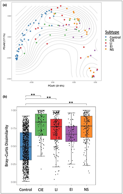
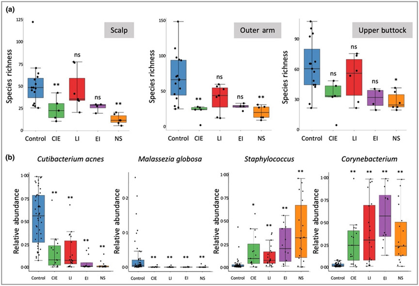
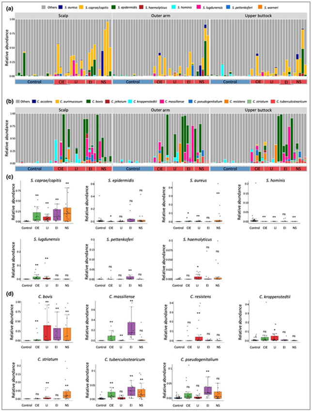
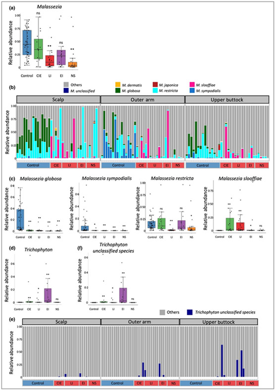
Results: Taxonomic profiling showed significant shifts in bacteria and fungi abundance and sporadic viral increases across ichthyosis subtypes. Cutibacterium acnes and Malassezia were significantly reduced across body sites, consistent with skin barrier disruption and depletion of lipids. Microbial richness was reduced, with specific increases in Staphylococcus and Corynebacterium genera, as well as shifts in fungal species, including Malassezia. Malassezia globosa was reduced at all body sites, whereas M. sympodialis was reduced in the ichthyotic upper arm and upper buttocks. Malassezia slooffiae, by contrast, was strikingly increased at all body sites in participants with congenital ichthyosiform erythroderma (CIE) and lamellar ichthyosis (LI). A previously undescribed Trichophyton species was also detected as sporadically colonizing the skin of patients with CIE, LI and epidermolytic ichthyosis subtypes.

Conclusions: The ichthyosis skin microbiome is significantly altered from healthy skin with specific changes predominating among ichthyosis subtypes. Skewing towards the Th17 pathway may represent a response to the altered microbial colonization in ichthyosis. What is already known about this topic? The skin microbiome of congenital ichthyoses is largely unexplored. Microbes play an important role in pathogenesis, as infections are common. The relative abundances of staphylococci and corynebacteria is increased in the cutaneous microbiome of patients with Netherton syndrome, but extension of these abundances to all congenital ichthyoses is unexplored. What does this study add? A common skin microbiome signature was observed across congenital ichthyoses. Distinct microbiome features were associated with ichthyosis subtypes. Changes in microbiome may contribute to T helper 17 cell immune polarization. What is the translational message? These data provide the basis for comparison of the microbiome with lipidomic and transcriptomic alterations in these forms of ichthyosis and consideration of correcting the dysbiosis as a therapeutic intervention.


Conflict of interest statement

Conflicts of interest

The authors declare they have no conflicts of interest.

Figures

Figure 1
Figure 1
Distinct skin microbiome community structures in congenital ichthyosis. (a) Bray–Curtis dissimilarity species-level analysis from multiple body sites. Principal coordinates analysis (PCoA) plot with contours to indicate sample density. (b) Box plots showing the Bray–Curtis dissimilarity of taxonomic profiles within control participants and within participants with ichthyosis subtypes. Statistical significance was determined using the permutation test on the Wilcoxon rank sum test in 100 000 times of random reshuffling of the participant subgroup labels with Bonferroni multiple testing correction (**P < 0·01; as described in the Method). CIE, congenital ichthyosiform erythroderma; EI, epidermolytic ichthyosis; LI, lamellar ichthyosis; NS, Netherton syndrome.
Figure 2
Figure 2
Major shifts in key community members in control and ichthyosis subtype skin. (a) Species richness is reduced in congenital ichthyosiform erythroderma (CIE) and Netherton syndrome (NS) on the scalp and the outer arm, and NS on the upper buttock. Kruskal–Wallis tests for three body sites are all significant (P-value of 3·22 × 10−4, 8·20 × 10−4 and 8·42 × 10−3 respectively for scalp, outer arm and upper buttock) and Bonferroni adjusted P-values from Dunn’s post hoc tests were used to indicate the statistical significance in comparison of subtypes vs. the control group (*P < 0·05; **P < 0·01). Raw and adjusted P-values can be found in Table S8; see Supporting Information. (b) Major significant changes in key microbial species were observed in all subtypes, including a reduction of Cutibacterium acnes and Malassezia globosa, and an increase in species in the Staphylococcus and Corynebacterium genera. Statistical significance was determined using the permutation test on the Wilcoxon rank sum test statistic with multiple testing correction using familywise error rate as described in Appendix S1 (see Supporting Information) (*P < 0·05; **P < 0·01). EI, epidermolytic ichthyosis; LI, lamellar ichthyosis; ns, not significant.
Figure 3.
Figure 3.
Stacked bar plots showing relative abundance of (a) Staphylococcus and (B) Corynebacterium spp. (P < 0·05) present in the healthy control and the congenital ichthyosis skin microbiome and grouped by body site. Healthy controls are labelled on the x-axis as control in the blue box and participants with ichthyosis are grouped according to disease subtype [congenital ichthyosiform erythroderma (CIE); lamellar ichthyosis (LI); epidermolytic ichthyosis (EI) and Netherton syndrome (NS)]. Box plots showing significant changes in relative abundance of (c) Staphylococcus and (d) Corynebacterium spp. In the ichthyosis subtypes in comparison with the healthy control group. Statistical significance was determined using the permutation test on the Wilcoxon rank sum test statistic with multiple testing correction using familywise error rate as described in Appendix S1 (see Supporting Information) (*P < 0·05; **P < 0·01) was computed for (c) and (d). ns, not significant.
Figure 4
Figure 4
Multikingdom analysis of skin microbiome. Multikingdom analysis plot from MetaPhlAn2 analysis shows relative abundance of (a) bacteria, increased in the participants with ichthyosis subtypes lamellar ichthyosis (LI); epidermolytic ichthyosis (EI) and Netherton syndrome (NS). (b) Eukaryota are significantly reduced in participants with ichthyosis subtypes compared with the healthy control group. (c) The viral kingdom analysis plot shows that the level of viruses on the skin of participants with ichthyosis is low, except for patients with congenital ichthyosiform erythroderma (CIE), who have a trend towards increased relative abundance at the three body sites that did not reach significance. The false discovery rate (FDR)-corrected Wilcoxon rank sum test P-value (*P < 0·05; **P < 0·01), undertaken separately for three body sites was used to determine the statistical significance. FDR multiple testing correction were undertaken over all the 12 tests of the three kingdoms and for the three body sites separately. Raw and adjusted P-values can be found in Table S8 (see Supporting Information). ns, not significant.
Figure 5
Figure 5
Mycobiome analysis for the control and ichthyosis groups. (a) Malassezia genus level alterations separated for ichthyosis subtypes. (b) Stacked bar charts of mycobiome highlighting only Malassezia spp. (P < 0·05) across the spectrum of ichthyosis subtypes. (c) Malassezia globosa and M. sympodialis were significantly reduced in all ichthyosis subtypes, whereas M. slooffiae was significantly increased in ichthyosis skin [except Netherton syndrome (NS)]. Malassezia restricta was significantly reduced in lamellar ichthyosis (LI) and NS subtypes. (d) Trichophyton at the genus level was augmented in all subtypes (except NS). (e) Stacked bar charts of mycobiome highlighting only Trichophyton spp. (P < 0·05) across the spectrum of ichthyosis subtypes. (f) Trichophyton unclassified species was significantly increased in all subtypes (except NS). Statistical significance was determined using permutation test on the Wilcoxon rank sum test statistic with multiple testing correction using familywise error rate as described in Appendix S1 (see Supporting Information) (*P < 0·05; **P < 0·01) was computed for (a), (c), (d) and (f). CIE, congenital ichthyosiform erythroderma; epidermolytic ichthyosis (EI); ns, not significant.

Comment in

References

    1. Oji V, Tadini G, Akiyama M et al. Revised nomenclature and classification of inherited ichthyoses: results of the First Ichthyosis Consensus Conference in Soreze 2009. J Am Acad Dermatol 2010; 63:607–41. - PubMed
    1. Vahlquist A, Fischer J, Torma H. Inherited nonsyndromic ichthyoses: an update on pathophysiology, diagnosis and treatment. Am J Clin Dermatol 2018; 19:51–66. - PMC - PubMed
    1. Brown SJ, Relton CL, Liao H et al. Filaggrin haploinsufficiency is highly penetrant and is associated with increased severity of eczema: further delineation of the skin phenotype in a prospective epidemiological study of 792 school children. Br J Dermatol 2009; 161:884–9. - PMC - PubMed
    1. Craig WY, Roberson M, Palomaki GE et al. Prevalence of steroid sulfatase deficiency in California according to race and ethnicity. Prenat Diagn 2010; 30:893–8. - PubMed
    1. Chavanas S, Bodemer C, Rochat A et al. Mutations in SPINK5, encoding a serine protease inhibitor, cause Netherton syndrome. Nat Genet 2000; 25:141–2. - PubMed