

Robustness of radiomic features in magnetic resonance imaging for patients with glioblastoma: Multi-center study
- PMID: 35633866
- PMCID: PMC9130546
- DOI: 10.1016/j.phro.2022.05.006
Robustness of radiomic features in magnetic resonance imaging for patients with glioblastoma: Multi-center study
Erratum in
-
Corrigendum to "Robustness of radiomic features in magnetic resonance imaging for patients with glioblastoma: Multi-center study" [Phys. Imaging Radiat. Oncol. 22 (2022) 131-136].Phys Imaging Radiat Oncol. 2022 Jun 23;23:43. doi: 10.1016/j.phro.2022.06.006. eCollection 2022 Jul. Phys Imaging Radiat Oncol. 2022. PMID: 35783579 Free PMC article.
Abstract
Background and purpose: Radiomics offers great potential in improving diagnosis and treatment for patients with glioblastoma multiforme. However, in order to implement radiomics in clinical routine, the features used for prognostic modelling need to be stable. This comprises significant challenge in multi-center studies. The aim of this study was to evaluate the impact of different image normalization methods on MRI features robustness in multi-center study.
Methods: Radiomics stability was checked on magnetic resonance images of eleven patients. The images were acquired in two different hospitals using contrast-enhanced T1 sequences. The images were normalized using one of five investigated approaches including grey-level discretization, histogram matching and z-score. Then, radiomic features were extracted and features stability was evaluated using intra-class correlation coefficients. In the second part of the study, improvement in the prognostic performance of features was tested on 60 patients derived from publicly available dataset.
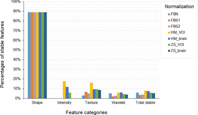
Results: Depending on the normalization scheme, the percentage of stable features varied from 3.4% to 8%. The histogram matching based on the tumor region showed the highest amount of the stable features (113/1404); while normalization using fixed bin size resulted in 48 stable features. The histogram matching also led to better prognostic value (median c-index increase of 0.065) comparing to non-normalized images.
Conclusions: MRI normalization plays an important role in radiomics. Appropriate normalization helps to select robust features, which can be used for prognostic modelling in multicenter studies. In our study, histogram matching based on tumor region improved both stability of radiomic features and their prognostic value.
Keywords: Features stability; Glioblastoma multiforme; Image normalization; Prognostic modelling; Radiomics.
© 2022 The Authors. Published by Elsevier B.V. on behalf of European Society of Radiotherapy & Oncology.
Conflict of interest statement
The authors declare that they have no known competing financial interests or personal relationships that could have appeared to influence the work reported in this paper.
Figures
References
-
- Weller M., van den Bent M., Hopkins K., Tonn J.C., Stupp R., Falini A., et al. EANO guideline for the diagnosis and treatment of anaplastic gliomas and glioblastoma. Lancet Oncol. 2014;15:e395–e403. - PubMed
-
- Li H., Li J., Cheng G., Zhang J., Li X. IDH mutation and MGMT promoter methylation are associated with the pseudoprogression and improved prognosis of glioblastoma multiforme patients who have undergone concurrent and adjuvant temozolomide-based chemoradiotherapy. Clin Neurol Neurosurg. 2016;151:31–36. - PubMed
-
- Bogowicz M., Vuong D., Huellner M.W., Pavic M., Andratschke N., Gabrys H.S., et al. CT radiomics and PET radiomics: ready for clinical implementation? Q J Nucl Med Mol Imaging. 2019;63:355–370. - PubMed
-
- Le Fevre C., Lhermitte B., Ahle G., Chambrelant I., Cebula H., Antoni D., et al. Pseudoprogression versus true progression in glioblastoma patients: A multiapproach literature review: Part 1 - Molecular, morphological and clinical features. Crit Rev Oncol Hematol. 2021;157 - PubMed
LinkOut - more resources
Full Text Sources
