

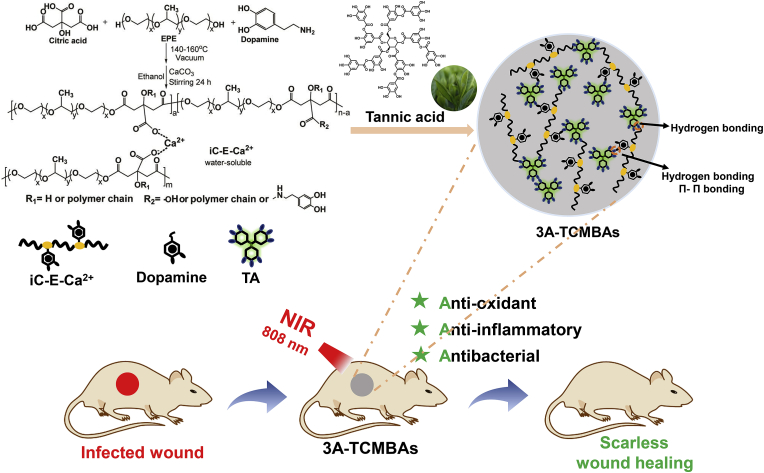
Anti-oxidant anti-inflammatory and antibacterial tannin-crosslinked citrate-based mussel-inspired bioadhesives facilitate scarless wound healing
- PMID: 35633874
- PMCID: PMC9131258
- DOI: 10.1016/j.bioactmat.2022.05.017
Anti-oxidant anti-inflammatory and antibacterial tannin-crosslinked citrate-based mussel-inspired bioadhesives facilitate scarless wound healing
Abstract
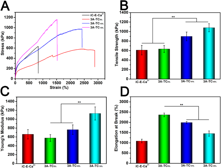
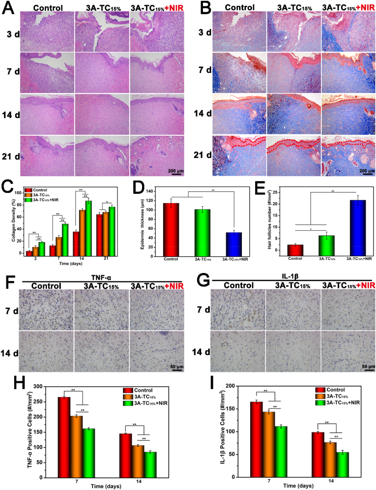
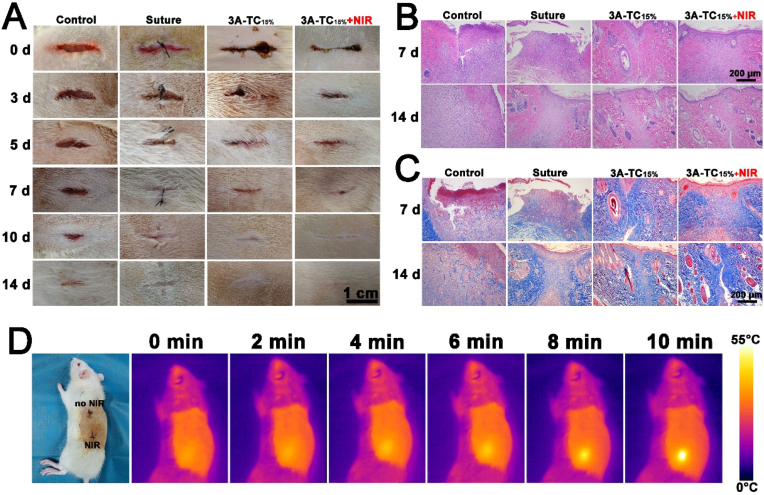
The revolutionary role of tissue adhesives in wound closure, tissue sealing, and bleeding control necessitates the development of multifunctional materials capable of effective and scarless healing. In contrast to the use of traditionally utilized toxic oxidative crosslinking initiators (exemplified by sodium periodate and silver nitrate), herein, the natural polyphenolic compound tannic acid (TA) was used to achieve near instantaneous (<25s), hydrogen bond mediated gelation of citrate-based mussel-inspired bioadhesives combining anti-oxidant, anti-inflammatory, and antimicrobial activities (3A-TCMBAs). The resulting materials were self-healing and possessed low swelling ratios (<60%) as well as considerable mechanical strength (up to ∼1.0 MPa), elasticity (elongation ∼2700%), and adhesion (up to 40 kPa). The 3A-TCMBAs showed strong in vitro and in vivo anti-oxidant ability, favorable cytocompatibility and cell migration, as well as photothermal antimicrobial activity against both Staphylococcus aureus and Escherichia coli (>90% bacterial death upon near-infrared (NIR) irradiation). In vivo evaluation in both an infected full-thickness skin wound model and a rat skin incision model demonstrated that 3A-TCMBAs + NIR treatment could promote wound closure and collagen deposition and improve the collagen I/III ratio on wound sites while simultaneously inhibiting the expression of pro-inflammatory cytokines. Further, phased angiogenesis was observed via promotion in the early wound closure phases followed by inhibition and triggering of degradation & remodeling of the extracellular matrix (ECM) in the late stage (supported by phased CD31 (platelet endothelial cell adhesion molecule-1) PDGF (platelet-derived growth factor) and VEGF (vascular endothelial growth factor) expression as well as elevated matrix metalloprotein-9 (MMP-9) expression on day 21), resulting in scarless wound healing. The significant convergence of material and bioactive properties elucidated above warrant further exploration of 3A-TCMBAs as a significant, new class of bioadhesive.
Keywords: Anti-oxidant; Hydrogen bond crosslinking; Phased angiogenesis; Scarless wound healing; Tannic acid.
© 2022 The Authors.
Conflict of interest statement
The authors declare that they have no known competing financial interests or personal relationships that could have appeared to influence the work reported in this paper.
Figures


References
-
- Zhou L., Zheng H., Liu Z., Wang S., Liu Z., Chen F., Zhang H., Kong J., Zhou F., Zhang Q. Conductive antibacterial hemostatic multifunctional scaffolds based on ti3c2tx mxene nanosheets for promoting multidrug-resistant bacteria-infected wound healing. ACS Nano. 2021;15(2):2468–2480. doi: 10.1021/acsnano.0c06287. - DOI - PubMed
-
- Luo M., Wang M., Niu W., Chen M., Cheng W., Zhang L., Xie C., Wang Y., Guo Y., Leng T. Injectable self-healing anti-inflammatory europium oxide-based dressing with high angiogenesis for improving wound healing and skin regeneration. Chem. Eng. J. 2021;412 doi: 10.1016/j.cej.2021.128471. - DOI
-
- Zhang S., Li Y., Qiu X., Jiao A., Luo W., Lin X., Zhang X., Zhang Z., Hong J., Cai P. Incorporating redox-sensitive nanogels into bioabsorbable nanofibrous membrane to acquire ROS-balance capacity for skin regeneration. Bioact. Mater. 2021;6(10):3461–3472. doi: 10.1016/j.bioactmat.2021.03.009. - DOI - PMC - PubMed
LinkOut - more resources
Full Text Sources
Miscellaneous
