

Early Diagnosis of Brain Tumour MRI Images Using Hybrid Techniques between Deep and Machine Learning
- PMID: 35633922
- PMCID: PMC9132638
- DOI: 10.1155/2022/8330833
Early Diagnosis of Brain Tumour MRI Images Using Hybrid Techniques between Deep and Machine Learning
Abstract
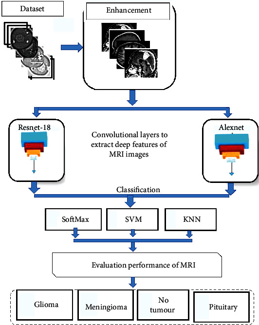
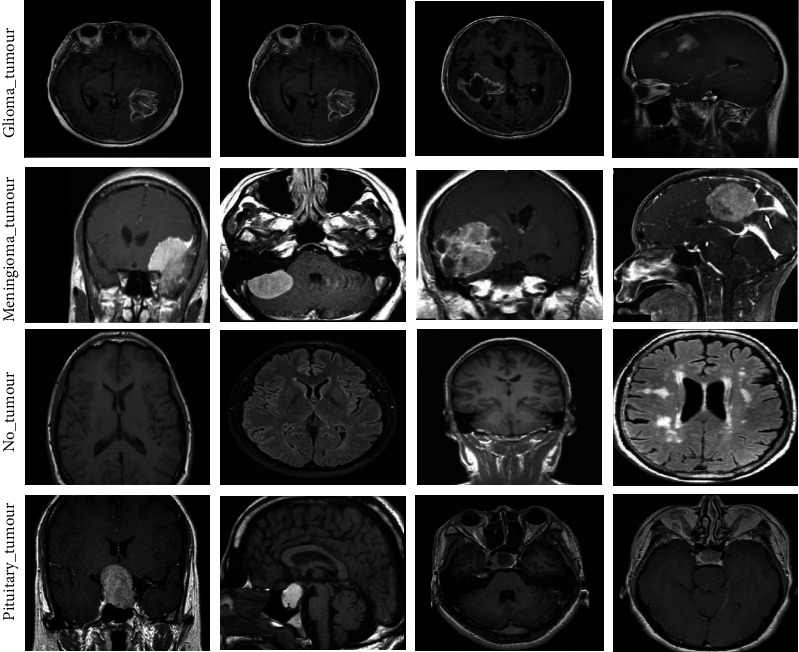
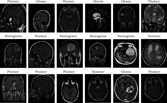
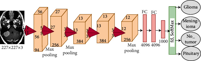
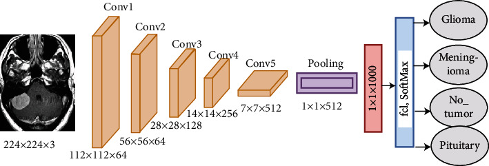
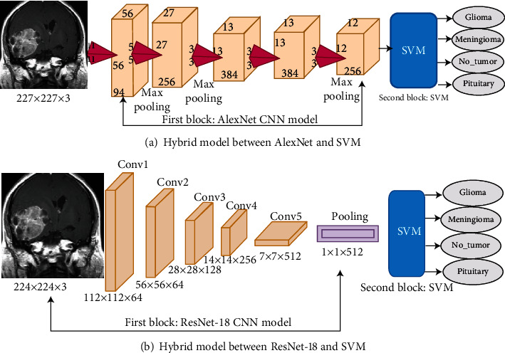
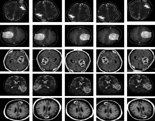
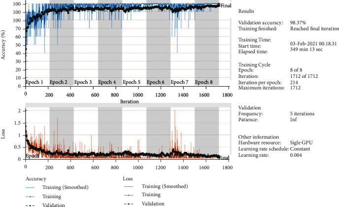
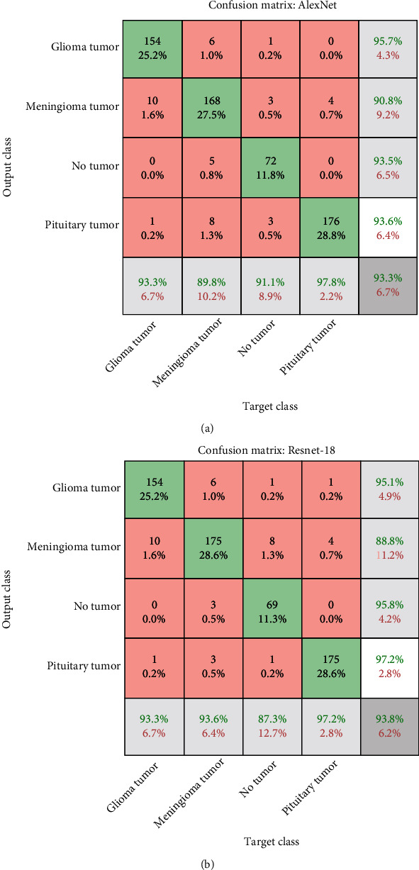
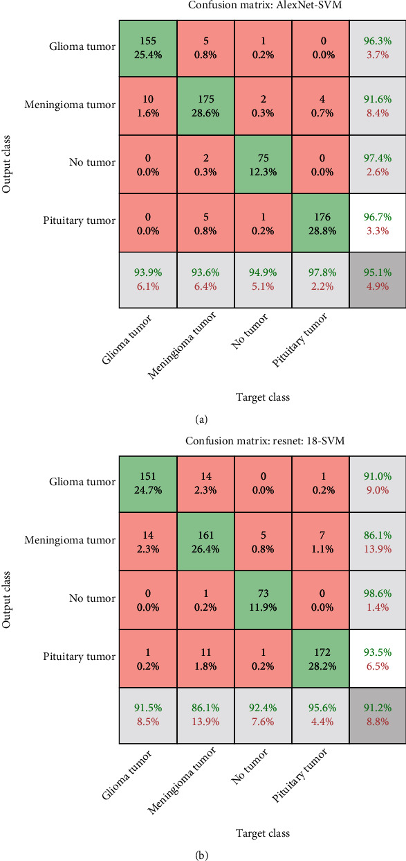
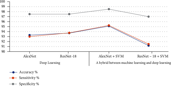
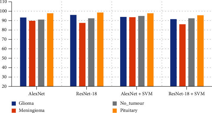
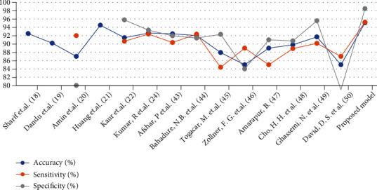
Cancer is considered one of the most aggressive and destructive diseases that shortens the average lives of patients. Misdiagnosed brain tumours lead to false medical intervention, which reduces patients' chance of survival. Accurate early medical diagnoses of brain tumour are an essential point for starting treatment plans that improve the survival of patients with brain tumours. Computer-aided diagnostic systems have provided consecutive successes for helping medical doctors make accurate diagnoses and have conducted positive strides in the field of deep and machine learning. Deep convolutional layers extract strong distinguishing features from the regions of interest compared with those extracted using traditional methods. In this study, different experiments are performed for brain tumour diagnosis by combining deep learning and traditional machine learning techniques. AlexNet and ResNet-18 are used with the support vector machine (SVM) algorithm for brain tumour classification and diagnosis. Brain tumour magnetic resonance imaging (MRI) images are enhanced using the average filter technique. Then, deep learning techniques are applied to extract robust and important deep features via deep convolutional layers. The process of combining deep and machine learning techniques starts, where features are extracted using deep learning techniques, namely, AlexNet and ResNet-18. These features are then classified using SoftMax and SVM. The MRI dataset contains 3,060 images divided into four classes, which are three tumours and one normal. All systems have achieved superior results. Specifically, the AlexNet+SVM hybrid technique exhibits the best performance, with 95.10% accuracy, 95.25% sensitivity, and 98.50% specificity.
Copyright © 2022 Ebrahim Mohammed Senan et al.
Conflict of interest statement
The authors declare no conflicts of interest regarding the publication of this paper.
Figures
References
-
- Roser M., Ritchie H. Cancer. 2019. https://ourworldindata.org/cancer .
-
- Swati Z. N. K., Zhao Q., Kabir M., et al. Content-based brain tumor retrieval for MR images using transfer learning. IEEE Access . 2019;7:17809–17822. doi: 10.1109/ACCESS.2019.2892455. - DOI
-
- Pereira S., Meier R., Alves V., Reyes M., Silva C. A. Understanding and Interpreting Machine Learning in Medical Image Computing Applications . Cham: Springer; 2018. Automatic brain tumor grading from MRI data using convolutional neural networks and quality assessment. - DOI
-
- Kumar S., Dabas C., Godara S. Classification of brain MRI tumor images: a hybrid approach. Procedia Computer Science . 2017;122:510–517. doi: 10.1016/j.procs.2017.11.400. - DOI
MeSH terms
LinkOut - more resources
Full Text Sources
Medical
