. 2022 May 12:13:880887.
doi: 10.3389/fimmu.2022.880887. eCollection 2022.

Macrophage Cx43 Is Necessary for Fibroblast Cytosolic Calcium and Lung Fibrosis After Injury

Affiliations

Macrophage Cx43 Is Necessary for Fibroblast Cytosolic Calcium and Lung Fibrosis After Injury

Aritra Bhattacharyya et al. Front Immunol. .

Abstract

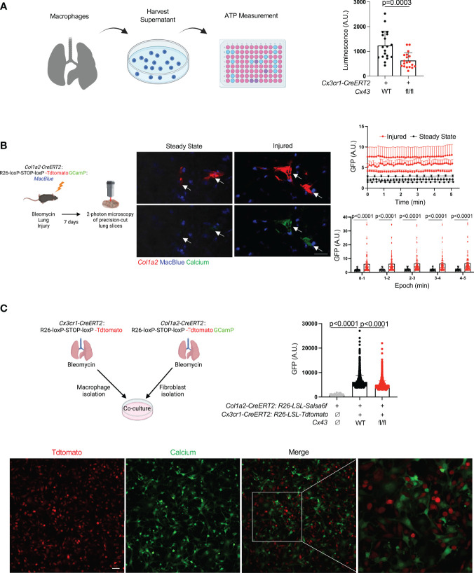
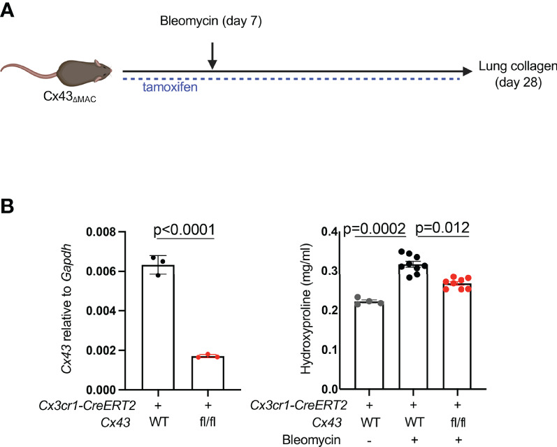
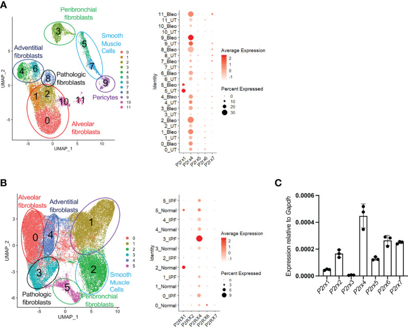
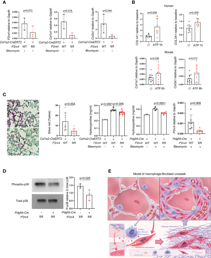
Macrophages are paracrine signalers that regulate tissular responses to injury through interactions with parenchymal cells. Connexin hemichannels have recently been shown to mediate efflux of ATP by macrophages, with resulting cytosolic calcium responses in adjacent cells. Here we report that lung macrophages with deletion of connexin 43 (MacΔCx43) had decreased ATP efflux into the extracellular space and induced a decreased cytosolic calcium response in co-cultured fibroblasts compared to WT macrophages. Furthermore, MacΔCx43 mice had decreased lung fibrosis after bleomycin-induced injury. Interrogating single cell data for human and mouse, we found that P2rx4 was the most highly expressed ATP receptor and calcium channel in lung fibroblasts and that its expression was increased in the setting of fibrosis. Fibroblast-specific deletion of P2rx4 in mice decreased lung fibrosis and collagen expression in lung fibroblasts in the bleomycin model. Taken together, these studies reveal a Cx43-dependent profibrotic effect of lung macrophages and support development of fibroblast P2rx4 as a therapeutic target for lung fibrosis.

Keywords: P2RX4; calcium imaging; fibroblast; lung fibrosis; macrophage.


Conflict of interest statement

The authors declare that the research was conducted in the absence of any commercial or financial relationships that could be construed as a potential conflict of interest.

Figures

Figure 1
Figure 1
Lung moMac Cx43 is required for ATP efflux and fibroblast cytosolic calcium in co-culture. (A) ATP measured in conditioned media after 24 hours of culture of tdTomato+ cells sorted from tamoxifen-induced Cx3cr1-CreERT2: LSL-tdTomato mice with or without homozygous floxed Cx43 alleles. Quantitation is for n=3 mice in each group, and points in the graph represent all technical replicates. P value is for Student’s unpaired two-tailed t-test. (B) Representative images of two-photon calcium imaging of precision-cut lung slices taken from Col1a2-CreERT2: R26-LSL-Salsa6f(tdtomato-GCaMP): MacBlue mice at steady state or 7 days after bleomycin injury. Arrows indicate fibroblasts. Scale bar= 50μm. Plots are for time-lapse imaging of individual slices from n=3 mice per condition (10-18 individual cells per field of view for injured and 5-11 individual cells per field of view for steady-state slices). Upper plot shows mean cytosolic GFP fluorescence for individual fibroblasts (as marked by tdTomato) for each slice across time; lower plot presents the same data as mean GFP fluorescence for aggregate data within 5 consecutive 1-minute time periods. P values are for 2-way ANOVA followed Sidak’s multiple comparison test. (C) Confocal calcium imaging of macrophages sorted from tamoxifen-induced Cx3cr1-CreERT2: R26-LSL-tdtomato mice and co-cultured with fibroblasts isolated from tamoxifen-induced Col1a2-CreERT2: R26-LSL-Salsa6f(tdtomato-GCaMP) mice. A representative field of view is shown, and the right-most image magnifies the indicated area in the merge. Scale bar= 50μm. Quantitation is for co-culture with WT or Cx43 KO macrophages, or for fibroblasts alone, for n=3 mice in each condition, and points in the graph represent individual cells. P values are for one-way ANOVA followed by Sidak’s multiple comparison test.
Figure 2
Figure 2
MoMac Cx43 is required for lung fibrosis. (A) Schematic for testing the effect of macrophage-specific Cx43 deletion on lung collagen after bleomycin injury with Cx3cr1-CreERT2: Cx43 fl/fl mice and controls. (B) qPCR of Cx43 expression in sorted tdTomato+ macrophages from Cx3cr1-CreERT2: R26-LSL-tdtomato: Cx43 fl/fl mice and Cx3cr1-CreERT2: R26-LSL-tdtomato controls. N=3 mice per condition. P value is for unpaired one-tailed t-test (left). Hydroxyproline assay of whole lung collagen from Cx3cr1-CreERT2: Cx43 fl/fl mice and Cx3cr1-CreERT2 controls. N=4, 9, and 8 mice as shown. P value is for one-way ANOVA followed by Kruskal-Wallis multiple comparison test (right).
Figure 3
Figure 3
P2rx4 is the most highly expressed ionotropic ATP receptor in lung fibroblasts. (A) UMAP plot of mouse fibroblasts from Tsukui et al. Dot plot shows expression of ionotropic ATP receptors. (B) UMAP plot of human fibroblasts from Tsukui et al. Dot plot shows expression of ionotropic ATP receptors. (C) qPCR of ionotropic ATP receptors in primary mouse lung fibroblasts isolated at steady state. N=3 mice.
Figure 4
Figure 4
P2rx4 is necessary for lung fibrosis. (A) qPCR for collagen genes and P2rx4 in primary mouse lung fibroblasts isolated from mice with fibroblast-specific P2rx4 deletion. N=3 mice. P values are for unpaired one-tailed t-tests. (B) qPCR for collagen genes in human (upper) and mouse (lower) primary lung fibroblasts treated with ATPγS. N=3 separate individuals and n=3 separate mice. P values are for unpaired two-tailed t-tests. (C) Left: Sirius red staining of lungs at 21 days post bleomycin injury in Col1a2-CreERT2: P2rx4 fl/fl or Col1a2-CreERT2 controls. Representative images are shown. Quantification is for n=3 mice. Scale bar=50μm. P value is for unpaired two-tailed t-test. Right: Hydroxyproline assays of whole lung collagen from mice with fibroblast-specific P2rx4 deletion with two different Cre drivers as indicated, and controls. N=3-10 as shown. P values are for one-way ANOVA followed by Sidak’s multiple comparison test. qPCR is for P2rx4 in fibroblasts isolated from Pdgfrb-Cre: P2rx4 fl/fl or Pdgfrb-Cre controls. N=6 mice in each group and P value is for Mann-Whitney test. (D) Western Blot of phospho-p38 and total p38 for fibroblasts isolated from Pdgfrb-Cre: P2rx4 fl/fl or Pdgfrb-Cre controls. Quantification shows the ratio of P-p38 to total p38 by densitometry. P value is shown for unpaired one-tailed t-test and n=3 mice. (E) Model of fibroblast activation by paracrine ATP derived from monocyte-derived macrophages in lung fibrosis.

References

    1. Misharin AV, Morales-Nebreda L, Reyfman PA, Cuda CM, Walter JM, Mcquattie-Pimentel AC, et al. . Monocyte-Derived Alveolar Macrophages Drive Lung Fibrosis and Persist in the Lung Over the Life Span. J Exp Med (2017) 214:2387–404. doi: 10.1084/jem.20162152 - DOI - PMC - PubMed
    1. Satoh T, Nakagawa K, Sugihara F, Kuwahara R, Ashihara M, Yamane F, et al. . Identification of an Atypical Monocyte and Committed Progenitor Involved in Fibrosis. Nature (2017) 541:96–101. doi: 10.1038/nature20611 - DOI - PubMed
    1. Aran D, Looney AP, Liu L, Wu E, Fong V, Hsu A, et al. . Reference-Based Analysis of Lung Single-Cell Sequencing Reveals a Transitional Profibrotic Macrophage. Nat Immunol (2019) 20:163–72. doi: 10.1038/s41590-018-0276-y - DOI - PMC - PubMed
    1. Larson-Casey JL, Deshane JS, Ryan AJ, Thannickal VJ, Carter AB. Macrophage Akt1 Kinase-Mediated Mitophagy Modulates Apoptosis Resistance and Pulmonary Fibrosis. Immunity (2016) 44:582–96. doi: 10.1016/j.immuni.2016.01.001 - DOI - PMC - PubMed
    1. Zhou X, Franklin RA, Adler M, Jacox JB, Bailis W, Shyer JA, et al. . Circuit Design Features of a Stable Two-Cell System. Cell (2018) 172:744–57.e717. doi: 10.1016/j.cell.2018.01.015 - DOI - PMC - PubMed

Publication types