. 2022 May 20:2022:7475704.
doi: 10.1155/2022/7475704. eCollection 2022.

Expression, Purification, and In Silico Characterization of Mycobacterium smegmatis Alternative Sigma Factor SigB

Affiliations

Expression, Purification, and In Silico Characterization of Mycobacterium smegmatis Alternative Sigma Factor SigB

Rakesh Kumar Singh et al. Dis Markers. .

Abstract

Sigma factor B (SigB), an alternative sigma factor (ASF), is very similar to primary sigma factor SigA (σ 70) but dispensable for growth in both Mycobacterium smegmatis (Msmeg) and Mycobacterium tuberculosis (Mtb). It is involved in general stress responses including heat, oxidative, surface, starvation stress, and macrophage infections. Despite having an extremely short half-life, SigB tends to operate downstream of at least three stress-responsive extra cytoplasmic function (ECF) sigma factors (SigH, SigE, SigL) and SigF involved in multiple signaling pathways. There is very little information available regarding the regulation of SigB sigma factor and its interacting protein partners. Hence, we cloned the SigB gene into pET28a vector and optimized its expression in three different strains of E. coli, viz., (BL21 (DE3), C41 (DE3), and CodonPlus (DE3)). We also optimized several other parameters for the expression of recombinant SigB including IPTG concentration, temperature, and time duration. We achieved the maximum expression of SigB at 25°C in the soluble fraction of the cell which was purified by affinity chromatography using Ni-NTA and further confirmed by Western blotting. Further, structural characterization demonstrates the instability of SigB in comparison to SigA that is carried out using homology modeling and structure function relationship. We have done protein-protein docking of RNA polymerase (RNAP) of Msmeg and SigB. This effort provides a platform for pulldown assay, structural, and other studies with the recombinant protein to deduce the SigB interacting proteins, which might pave the way to study its signaling networks along with its regulation.


Conflict of interest statement

The authors declare no conflict of interest.

Figures

Figure 1
Figure 1
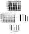
Expression host strain optimization for the expression of recombinant His-SigB. 10% SDS PAGE depicting recombinant His-SigB protein expression profile from three expression host strains, namely, BL21(DE3), C41 (DE3), and CodonPlus (DE3) expressed with 0.1 mM IPTG grown for 3 hrs at 37°C with shaking at 150 rpm. The highest protein expression is observed in the host strain CodonPlus (DE3). M: molecular size marker; V.O.: pET28a vector only control; U: uninduced His-SigB clone; I: induced His-SigB clone.
Figure 2
Figure 2
IPTG concentration optimization for the expression of recombinant His-SigB. (a) 10% SDS PAGE depicting recombinant His-SigB protein expression profile in CodonPlus (DE3) host strain. The highest protein expression is observed in 0.1 mM IPTG. (b) Solubility of the recombinant His-SigB in different IPTG concentrations at 37°C. 10% SDS PAGE depicting recombinant His-SigB protein distribution in cell pellet and supernatant in CodonPlus (DE3) induced with IPTG concentration from 0.05 to 0.5 mM IPTG. The highest protein solubility observed in the supernatant fraction at 0.1 mM IPTG. M: molecular size marker; V.O.: pET28a vector only control; V.O.I: pET28a vector only induced; U: uninduced His-SigB clone; P: pellet; S: supernatant. (c) Gel-based semiquantitative analysis from (a) also depicts the highest expression of His-SigB at 0.1 mM IPTG concentration. (d) Semiquantitative analysis from (b) depicts higher relative protein solubility observed in the supernatant fraction at 0.1 mM IPTG. Semiquantitative analysis of the gels was performed with Quantity One software (BIO-RAD).
Figure 3
Figure 3
Temperature optimization for the expression of recombinant His-SigB protein. (a) 10% SDS PAGE depicting recombinant His-SigB protein expression profile in CodonPlus (DE3) cells at temperatures: 37°C, 25°C, and 16°C with 0.1 mM IPTG. The highest protein expression was observed at 37°C. (b) 10% SDS PAGE depicting distribution of recombinant His-SigB protein in cell pellet and supernatant fractions at different temperatures. The highest protein solubility was observed at 25°C supernatant fraction. M: marker; V.O.: pET28a vector only control; U: uninduced His-SigB clone; P: pellet; S: supernatant. (c) Gel-based semiquantitative analysis from (a) depicts the highest expression of His-SigB at 37°C with 0.1 mM IPTG concentration. (d) Semiquantitative analysis from (b) depicts higher relative protein solubility observed in the supernatant fraction at 25°C with 0.1 mM IPTG. Semiquantitative analysis of the gels was performed with Quantity One software (BIO-RAD).
Figure 4
Figure 4
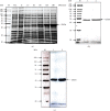
(a) The expression of recombinant His-SigB protein at different time points. 10% SDS PAGE depicting recombinant His-SigB protein expression profile collected at time points 15, 30, 60, 120, and 180 minutes. The highest protein expression was observed at 180 minutes after induction with 0.1 mM IPTG. (b) Ni-NTA affinity purification of His-SigB: lanes denoting M: molecular size marker; 1: 5 μg; 2: 7.5 μg His-SigB purified protein stained with Coomassie R-250. (c) Western blotting with purified recombinant His-SigB. Mouse anti-His monoclonal antibody (Genetix) was used to probe purified His-SigB. M: marker; V.O.: pET28a vector only control; U: uninduced His-SigB clone; lane 1: 5 μg; lane 2: 10 μg purified His-SigB protein.
Figure 5
Figure 5
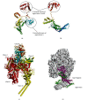
Homology models of (a) M. smegmatis sigma factor SigA, (b) M. smegmatis sigma factor SigB, (c) structure of the core RNA polymerase of M. smegmatis, and (d) comparative surface topology of SigA and SigB interactions with core RNA polymerase of M. smegmatis.

References

    1. Murakami K. S., Masuda S., Darst S. Structural basis of transcription initiation: RNA polymerase holoenzyme at 4 Å resolution. Science . 2002;296(5571):1280–1284. doi: 10.1126/science.1069594. - DOI - PubMed
    1. Vassylyev D. G., Sekine S. I., Laptenko O., et al. Crystal structure of a bacterial RNA polymerase holoenzyme at 2.6 Å resolution. Nature . 2002;417(6890):712–719. doi: 10.1038/nature752. - DOI - PubMed
    1. Manganelli R., Dubnau E., Tyagi S., Kramer F. R., Smith I. Differential expression of 10 sigma factor genes in Mycobacterium tuberculosis. Molecular Microbiology . 1999;31(2):715–724. doi: 10.1046/j.1365-2958.1999.01212.x. - DOI - PubMed
    1. Wu S., Howard S. T., Lakey D. L., et al. The principal sigma factor sigA mediates enhanced growth of Mycobacterium tuberculosis in vivo. Molecular Microbiology . 2004;51(6):1551–1562. doi: 10.1111/j.1365-2958.2003.03922.x. - DOI - PubMed
    1. Paget M. S. B., Helmann J. D. The σ70 family of sigma factors. Genome Biology . 2003;4(1):203–206. doi: 10.1186/gb-2003-4-1-203. - DOI - PMC - PubMed

MeSH terms

LinkOut - more resources