. 2022 Nov;53(6):543-557.
doi: 10.1177/15500594221103834. Epub 2022 May 29.

Changes in Electrical Brain Activity and Cognitive Functions Following Mild to Moderate COVID-19: A one-Year Prospective Study After Acute Infection

Affiliations

Changes in Electrical Brain Activity and Cognitive Functions Following Mild to Moderate COVID-19: A one-Year Prospective Study After Acute Infection

Pablo Andrei Appelt et al. Clin EEG Neurosci. 2022 Nov.

Abstract

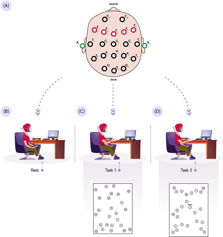
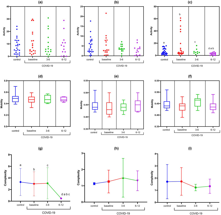
The coronavirus disease 2019 (COVID-19) can disrupt various brain functions. Over a one-year period, we aimed to assess brain activity and cognitive function in 53 COVID-19 patients and 30 individuals without COVID-19 (or asymptomatic). The Montreal Cognitive Assessment, Trail Making Test Parts A and B (TMT-A and B), and Digit Span Test were used to assess cognitive function. Cognitive variables and electroencephalography (EEG) data (activity, mobility, and complexity) were compared between the groups at rest and during cognitive demand (F3-F7, Fz-F3, Fz-F4, and F4-F8). There was a reduction in F3-F7 activity during the TMT-B in the COVID-19 group at 6-12 months compared to the controls (p = 0.01) at baseline (p = 0.03), a reduction in signal complexity at F3-F7 at rest in the COVID-19 group at baseline and 6-12 months compared to the controls (p < 0.001), and a reduction in Fz-F4 activity at rest from 6-12 months in the post-COVID group compared to baseline (p = 0.02) and 3-6 months (p = 0.04). At 6-12 months, there was a time increase in TMT-A in the COVID-19 group compared to that in the controls (p = 0.04). Some correlations were found between EEG data and cognitive test in both groups. In conclusion, there was a reduction in brain activity at rest in the Fz-F4 areas and during high cognitive demands in the F3-F7 areas. A reduction in signal complexity in F3-F7 at rest was found in the COVID-19 group at 6-12 months after acute infection. Furthermore, individuals with COVID-19 experience long-term changes in cognitive function.

Keywords: COVID-19; SARS-CoV-2; cognition; electroencephalography.


Conflict of interest statement

The author(s) declared no potential conflicts of interest with respect to the research, authorship, and/or publication of this article.

Figures

Figure 1.
Figure 1.
EEG protocol. (A): PFC EEG acquisition; (B) EEG acquisition at rest; (C) EEG acquisition during TMT-A (low cognitive demand); (D) EEG acquisition during TMT-B (high cognitive demand).
Figure 2.
Figure 2.
Statistical properties used in signal processing in the time domain of the F3-F7 channel in the control and COVID-19 groups. (A) F3-F7 activity at rest; (B) F3-F7 activity during TMT-A; (C) F3-F7 activity during TMT-B; (D) F3-F7 mobility at rest; (E) F3-F7 mobility during TMT-A; (F) F3-F7 mobility during TMT-B; (G) F3-F7 complexity at rest; (H) F3-F7 complexity during TMT-A; (I) F3-F7 complexity during TMT-A.
Figure 3.
Figure 3.
Statistical properties used in signal processing in the time domain of the Fz-F3 channel in the control and COVID-19 groups. (A) Fz-F3 activity at rest; (B) Fz-F3 activity during TMT-A; (C) Fz-F3 activity during TMT-B; (D) Fz-F3 mobility at rest; (E) Fz-F3 mobility during TMT-A; (F) Fz-F3 mobility during TMT-B; (G) Fz-F3 complexity at rest; (H) Fz-F3 complexity during TMT-A; (I) Fz-F3 complexity TMT-A.
Figure 4.
Figure 4.
Statistical properties used in signal processing in the time domain of the Fz-F4 channel in the control and COVID-19 groups. (A) Fz-F4 activity at rest; (B) Fz-F4 activity during TMT-A; (C) Fz-F4 activity during TMT-B; (D) Fz-F4 mobility at rest; (E) Fz-F4 mobility during TMT-A; (f) Fz-F4 mobility during TMT-B; (G) Fz-F4 complexity at rest; (H) Fz-F4 complexity during TMT-A; (I) Fz-F4 complexity during TMT-A.
Figure 5.
Figure 5.
Statistical properties used in time domain signal processing of the F4-F8 channel in the control and COVID-19 groups. (A) F4-F8 activity at rest; (b) F4-F8 activity during TMT-A; (c) F4-F8 activity during TMT-B; (d) F4-F8 mobility at rest; (e) F4-F8 mobility during TMT-A; (f) F4-F8 mobility during TMT-B; (g) F4-F8 complexity at rest; (h) F4-F8 complexity during TMT-A; (i) F4-F8 complexity during TMT-A.

References

    1. Li Q, Guan X, Wu Pet al.Early transmission dynamicsinWuhan,China,ofnovelcoronavirus- infected pneumonia. N Engl J Med. 2020;382(13):1199–1207. doi:10.1056/NEJMoa2001316 - DOI - PMC - PubMed
    1. Huang C, Wang Y, Li Xet al.Clinical features of patients infected with 2019 novel coronavirus in Wuhan, China. Lancet. 2020;395(10223):497–506. 10.1016/S0140-6736(20)30183-5 - DOI - PMC - PubMed
    1. Mao L, Jin H, Wang Met al.Neurologic manifestations of hospitalized patients with coronavirus disease 2019 in Wuhan, China. JAMA Neurol. 2020;77(6):683–690. https://.doi:10.1001/jamaneurol.2020.1127 - DOI - PMC - PubMed
    1. Crook H, Raza S, Nowell J, Young M, Edison P. Long COVID - mechanisms, risk factors, and management. Br Med J. 2021;37(4):n1648. 10.1136/bmj.n1648 - DOI - PubMed
    1. Romero-Sánchez CM, Díaz-Maroto I, Fernández-Díaz Eet al.Neurologic manifestations in hospitalized patients with COVID-19: the ALBACOVID registry. Neurology. 2020;95(8):e1060–e1070. doi:10.1212/WNL.0000000000009937 - DOI - PMC - PubMed