. 2022 Aug:62:101518.
doi: 10.1016/j.molmet.2022.101518. Epub 2022 May 28.

Proteasome dysfunction disrupts adipogenesis and induces inflammation via ATF3

Affiliations

Proteasome dysfunction disrupts adipogenesis and induces inflammation via ATF3

Nienke Willemsen et al. Mol Metab. 2022 Aug.

Abstract

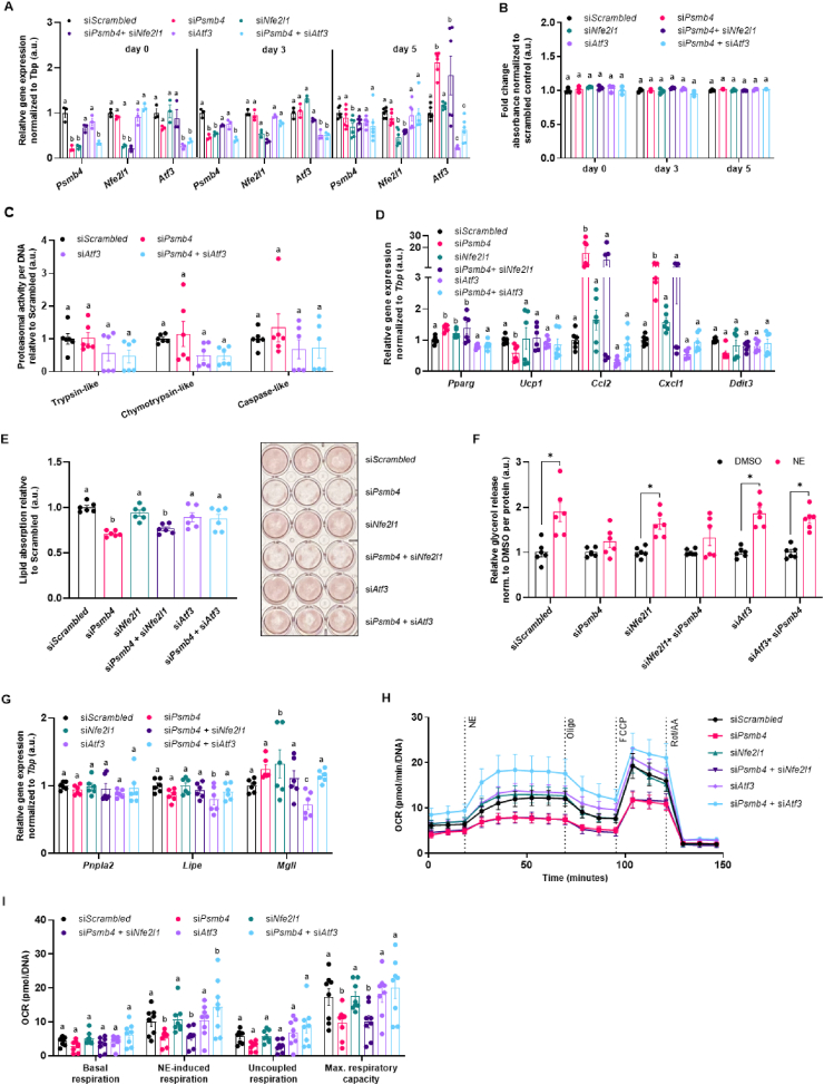
Objective: Regulation of proteasomal activity is an essential component of cellular proteostasis and function. This is evident in patients with mutations in proteasome subunits and associated regulators, who suffer from proteasome-associated autoinflammatory syndromes (PRAAS). These patients display lipodystrophy and fevers, which may be partly related to adipocyte malfunction and abnormal thermogenesis in adipose tissue. However, the cell-intrinsic pathways that could underlie these symptoms are unclear. Here, we investigate the impact of two proteasome subunits implicated in PRAAS, Psmb4 and Psmb8, on differentiation, function and proteostasis of brown adipocytes.

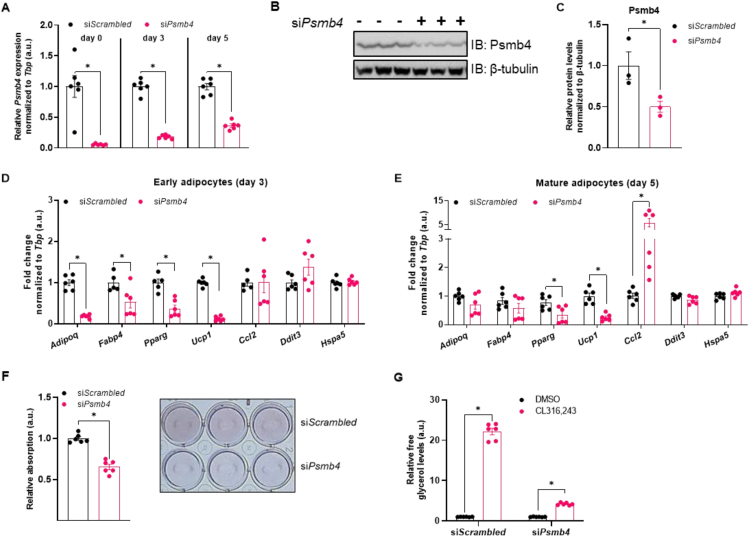
Methods: In immortalized mouse brown pre-adipocytes, levels of Psmb4, Psmb8, and downstream effectors genes were downregulated through reverse transfection with siRNA. Adipocytes were differentiated and analyzed with various assays of adipogenesis, lipogenesis, lipolysis, inflammation, and respiration.

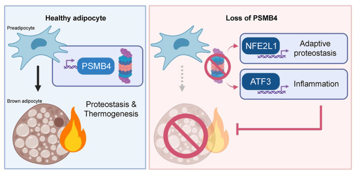
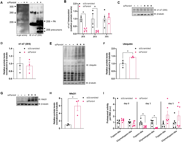
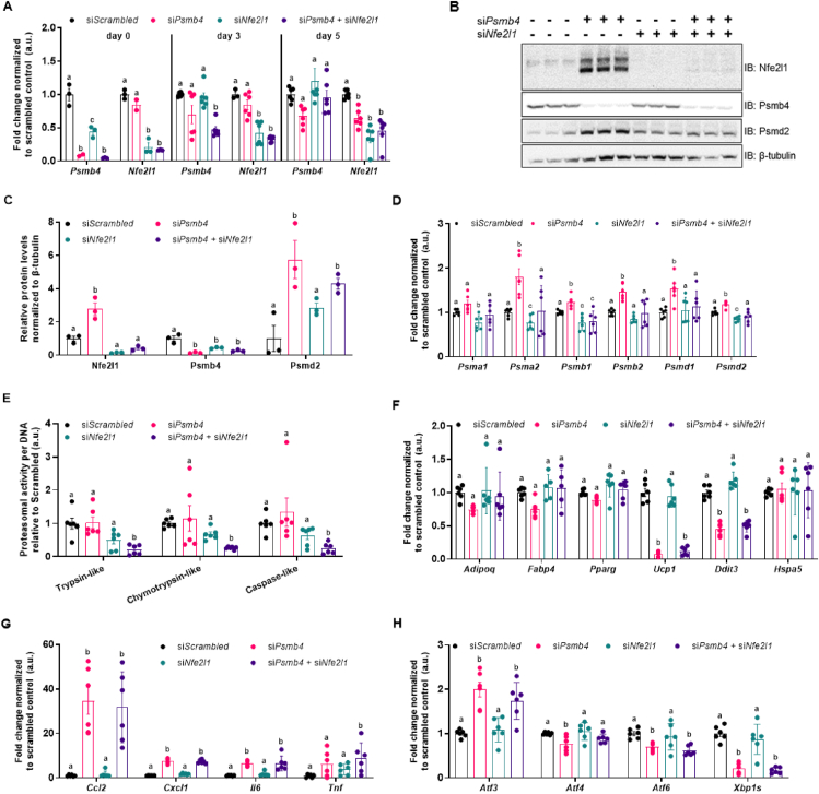
Results: Loss of Psmb4, but not Psmb8, disrupted proteostasis and adipogenesis. Proteasome function was reduced upon Psmb4 loss, but partly recovered by the activation of Nuclear factor, erythroid-2, like-1 (Nfe2l1). In addition, cells displayed higher levels of surrogate inflammation and stress markers, including Activating transcription factor-3 (Atf3). Simultaneous silencing of Psmb4 and Atf3 lowered inflammation and restored adipogenesis.

Conclusions: Our study shows that Psmb4 is required for adipocyte development and function in cultured adipocytes. These results imply that in humans with PSMB4 mutations, PRAAS-associated lipodystrophy is partly caused by disturbed adipogenesis. While we uncover a role for Nfe2l1 in the maintenance of proteostasis under these conditions, Atf3 is a key effector of inflammation and blocking adipogenesis. In conclusion, our work highlights how proteasome dysfunction is sensed and mitigated by the integrated stress response in adipocytes with potential relevance for PRAAS patients and beyond.

Keywords: ATF3; Adipocytes; NFE2L1; PSMB4; Proteasome; Proteostasis; Ubiquitin; brown adipose tissue.


Figures

Image 1
Graphical abstract
Figure 1
Figure 1
Psmb4 controls adipogenesis and adipocyte health. (A) Relative gene expression of Psmb4 at different time points after knockdown with scrambled or Psmb4 siRNA. (B,C) Representative immunoblot of Psmb4 in siScrambled and siPsmb4 adipocytes with (C) protein quantification normalized to β-tubulin. (D) Relative gene expression of adipogenesis and stress markers adipocytes after Psmb4 knockdown measured on day 3 of differentiation. (E) Relative gene expression of adipogenesis and stress markers adipocytes after Psmb4 knockdown measured on day 5 of differentiation. (F) Representative Oil-Red-O staining and quantification (day 5). (G) Relative free glycerol levels in adipocyte supernatant (day 5) after DMSO or 1 μM CL316,243 treatment for 3 h. Data are mean ± SEM. Unless otherwise specified: n = 6 independent measurements from 2 separate experiments. Significant if P < 0.05, indicated by (∗) or different letters.
Figure 2
Figure 2
Psmb4 controls UPS and proteostasis. (A) Representative Native PAGE with in-gel chymotrypsin-like proteasome activity and immunoblot of ⍺1-⍺7 (20S) proteasome subunits. (PA: proteasome activators) (B) Relative protein levels ⍺1-⍺7 (20S) proteasome subunits measured in Native PAGE immunoblots. (C,D) Representative immunoblot of ⍺1-⍺7 (20S) proteasome subunits from adipocytes (day 5) with (D) protein quantification relative to β-tubulin. (E,F) Representative immunoblot of ubiquitin from adipocytes (day 5) with (F) protein quantification relative to β-tubulin. (G,H) Representative immunoblot of Nfe2l1 from adipocytes (day 5) with (D) protein quantification relative to β-tubulin. (I) Trypsin-like and chymotrypsin-like proteasome activity in adipocytes at different time points, normalized to DNA content. Data are mean ± SEM. Unless otherwise specified: n = 6 independent measurements from 2 separate experiments. Significant if P < 0.05, indicated by (∗) or different letters.
Figure 3
Figure 3
Loss of Psmb4 initiates a proteostatic stress-response via Nfe2l1. (A) Relative gene expression of Psmb4 and Nfe2l1 at different time points after knockdown with scrambled, Psmb4 and/or Nfe2l1 siRNA. (B,C) Representative immunoblots of Nfe2l1, Psmb4, Psmd2 and β-tubulin in siScrambled, siPsmb4, siNfe2l1 or siPsmb4 + siNfe2l1 adipocytes (day 5) with (C) protein quantification levels normalized to β-tubulin.(D) Relative gene expression of various proteasome subunits. (E) Trypsin-like, chymotrypsin-like, and caspase-like proteasome activity in adipocytes (day 5), normalized to DNA content. (F) Relative gene expression of adipogenesis and stress markers in adipocytes (day 5). (G) Relative gene expression of inflammation markers in adipocytes (day 5). (H) Relative gene expression of ER stress and unfolded protein response markers in adipocytes (day 5). Data are mean ± SEM. Unless otherwise specified: n = 6 independent measurements from 2 separate experiments. Significant if P < 0.05, indicated by (∗) or different letters.
Figure 4
Figure 4
Loss of Psmb4 induces inflammation and blocks adipogenesis via Atf3. (A) Relative gene expression of Psmb4, Nfe2l1, and Atf3. (B) Viability in adipocytes. (C) Trypsin-like, chymotrypsin-like, and caspase-like proteasome activity in adipocytes (day 5), normalized to DNA content (D) Relative gene expression of adipogenesis and stress markers in adipocytes (day 5) after knockdown. (E) Oil-Red-O staining in adipocytes after knockdown. (F) Supernatant free glycerol levels after treatment with DMSO or 1 μM Norepinephrine (NE) for 1 h normalized to protein. (G) Relative gene expression of lipases in adipocytes (day 5). (H, I) Oxygen consumption rate (OCR) in adipocytes after knockdown, normalized to DNA levels (n = 8, from 2 experiments). Unless otherwise specified: n = 6 independent measurements from 2 separate experiments. Data are mean ± SEM. Significant if P < 0.05, indicated by (∗) or different letters.

Similar articles

Cited by

References

    1. Torrelo A., Patel S., Colmenero I., Gurbindo D., Lendínez F., Hernández A., et al. Chronic atypical neutrophilic dermatosis with lipodystrophy and elevated temperature (CANDLE) syndrome. Journal of the American Academy of Dermatology. 2010 Mar;62(3):489–495. - PubMed
    1. Agarwal A.K., Xing C., DeMartino G.N., Mizrachi D., Hernandez M.D., Sousa A.B., et al. PSMB8 encoding the β5i proteasome subunit is mutated in Joint Contractures, muscle atrophy, microcytic anemia, and panniculitis-induced lipodystrophy syndrome. The American Journal of Human Genetics. 2010 Dec;87(6):866–872. - PMC - PubMed
    1. Kanazawa N. Nakajo-nishimura syndrome: an autoinflammatory disorder showing pernio-like rashes and progressive partial lipodystrophy. Allergology International. 2012;61(2):197–206. - PubMed
    1. Kitamura A., Maekawa Y., Uehara H., Izumi K., Kawachi I., Nishizawa M., et al. A mutation in the immunoproteasome subunit PSMB8 causes autoinflammation and lipodystrophy in humans. Journal of Clinical Investigation. 2011 Oct 3;121(10):4150–4160. - PMC - PubMed
    1. Brehm A., Liu Y., Sheikh A., Marrero B., Omoyinmi E., Zhou Q., et al. Additive loss-of-function proteasome subunit mutations in CANDLE/PRAAS patients promote type I IFN production. Journal of Clinical Investigation. 2015 Oct 20;125(11):4196–4211. - PMC - PubMed

Publication types

Substances