Review
. 2022 Sep;71(9):1892-1908.
doi: 10.1136/gutjnl-2021-326560. Epub 2022 May 30.

Organoids in gastrointestinal diseases: from experimental models to clinical translation

Affiliations
Review

Organoids in gastrointestinal diseases: from experimental models to clinical translation

Claudia Günther et al. Gut. 2022 Sep.

Abstract

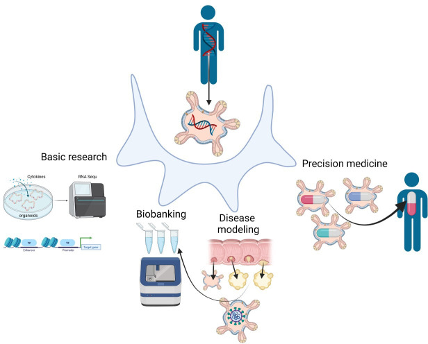
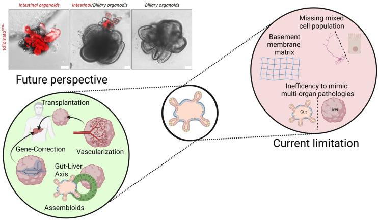
We are entering an era of medicine where increasingly sophisticated data will be obtained from patients to determine proper diagnosis, predict outcomes and direct therapies. We predict that the most valuable data will be produced by systems that are highly dynamic in both time and space. Three-dimensional (3D) organoids are poised to be such a highly valuable system for a variety of gastrointestinal (GI) diseases. In the lab, organoids have emerged as powerful systems to model molecular and cellular processes orchestrating natural and pathophysiological human tissue formation in remarkable detail. Preclinical studies have impressively demonstrated that these organs-in-a-dish can be used to model immunological, neoplastic, metabolic or infectious GI disorders by taking advantage of patient-derived material. Technological breakthroughs now allow to study cellular communication and molecular mechanisms of interorgan cross-talk in health and disease including communication along for example, the gut-brain axis or gut-liver axis. Despite considerable success in culturing classical 3D organoids from various parts of the GI tract, some challenges remain to develop these systems to best help patients. Novel platforms such as organ-on-a-chip, engineered biomimetic systems including engineered organoids, micromanufacturing, bioprinting and enhanced rigour and reproducibility will open improved avenues for tissue engineering, as well as regenerative and personalised medicine. This review will highlight some of the established methods and also some exciting novel perspectives on organoids in the fields of gastroenterology. At present, this field is poised to move forward and impact many currently intractable GI diseases in the form of novel diagnostics and therapeutics.

Keywords: gastrointestinal pathology; gastrointestinal physiology; intestinal stem cell; stem cells.


Conflict of interest statement

Competing interests: None declared.

Figures

Figure 1
Figure 1
Organoids, assembly of single cells. The term organoid generally applies to three-dimensional cellular cultures derived from stem cells that resemble components of a complex tissue or organ.
Figure 2
Figure 2
The GI organoid culture system. GI organoids can be derived from PSCs and ASCs. PSC-derived organoids are established by directed differentiation of either ESCs or iPSCs (generated from fibroblast of skin biopsies) towards the endoderm. ASCs of epithelial origin can be isolated from tissue biopsies or surgical resections. They can be embedded in ECM and grown from either single cells or crypts that give rise to organoids that contain crypt-like structures. Adaption of the culture medium can foster the development of spheroids that are enriched for stem cells. Depending on the culture condition both PSCs and ASCs can give rise to three-dimensional organ-like structures of the hepatic-biliary-pancreatic tract and digestive tract containing gastric, small intestinal and colonic organoids. Representative pictures of organoid cultures are derived from the authors (unpublished, scale bars: 50 µm). ASCs, adult stem cells; ESCs, embryonic stem cells; ECM, extracellular matrices; PSCs, pluripotent stem cells.
Figure 3
Figure 3
Overview of organoid derivation. GI organoids can be either derived from ASCs (left panel) or PSCs (right panel). Left panel: tissue-resident stem cells can be isolated from GI biopsies collected during colonoscopy, gastroscopy or surgery. Following an ethylenediaminetetraacetic acid (EDTA) or enzymatic digestion, either single cells or tissue fragments (crypt structures or ductal fragments) are embedded into an ECM and covered with a defined culture medium supplemented with a combination of different mitogens, morphogens and cytokines that guide the self-renewal and organised differentiation of somatic stem cells. Beside ASCs, organoids can be generated from cancer stem cells (isolated from tumour biopsies) and are referred to as tumouroids. Right panel: GI organoids can be also derived from PSCs (ESCs and iPSCs) in a stepwise differentiation process that recapitulates the signalling programme active during development. In an initial step, PSCs follow a guided differentiation towards an endodermal fate by exposure to Activin A. This is followed by the differentiation towards foregut, midgut/hindgut endoderm depending on the targeted tissue (foregut for gastric, hepatic, biliary or pancreatic organoids; midgut/hindgut for intestinal organoids). These precursor spheroids can be further instructed towards defined tissues and organs depending on different growth factors that allow proper terminal differentiation. ASCs, adult stem cells; CRC, colorectal cancer; ECM, extracellular matrices; ESCs, embryonic stem cells; IBD, inflammatory bowel disease; iPSCs, induced pluripotent stem cells.
Figure 4
Figure 4
Organoid coculture applications to increase the complexity of GI organoids, cellular and microbial components can be integrated into organoid cultures. This can be achieved by several protocols. (A) Reversal of the polarity in combination with removal of the ECM results in organoid cultures in which the apical/luminal surface faces towards the medium (Apical-out). (B) 3D-derived submerged monolayers can be grown as a thin monolayer to study the interaction of epithelial cells with environmental factors. (C) This monolayer technique has been expanded to air–liquid interface cultures in which monolayers are plated on transwells. This allows the investigation of the tissue microenvironment by adding defined cell populations (eg, immune cells, fibroblasts, endothelial cells) to the basal side of the epithelium and environmental factors to the apical surface. (D) Microinjections of luminal factors (eg, bacteria, viruses, dietary factors, metabolites) allow the investigation of the effect of environmental elements on epithelial homeostasis without changing architecture or polarity of organoids. (E) The organoid-on-a-chip concept is a more complex culture method in which self-organisation of stem cells can be extrinsically guided by a 3D microstructured scaffold. The advantage of this method is to integrate several cell types and mechano-physiological parameters. Asterisks indicate crypt region; L: lumen; red arrows indicate luminal side; air–liquid interface (C): pictures are derived from Wang et al., Cell 2019, Other representative pictures of organoid cultures are derived from the authors (unpublished, scale bars: 50 µm). 3D, three-dimensional; ECM, extracellular matrices.
Figure 5
Figure 5
Applications of organoids from patients can be used for basic research to better understand organ development and to dissect the relevant intracellular pathways that control homeostasis and might be altered during organ dysfunction. These patient-derived organoids can be further used to preclinically screen for novel therapies and to predict response to drugs as well as for regenerative medicine (precision medicine). Another preclinical application includes disease modelling to better understand the mechanisms underlying GI diseases (including inflammatory, infectious, metabolic, inheritable genetic and neoplastic disorders) as well as the biobanking of patient-derived-organoids and -tumouroids.
Figure 6
Figure 6
Current limitations and future perspective of GI organoids. Current limitations of GI organoids include their inability to model multiorgan pathologies and their limited cellular diversity. Assembloids (organoids derived from spatially organising multiple cell types) will partially address these limitations and allow us in the future to better understand cell-to-cell communication and organ-cross-talk. Finally the mechanistic impact of the EMC on organoids and the batch-to-batch variations are limiting factors for translational approaches. Several attempts have been made to replace the current ECM by natural or synthetic ECMs. Organoids were derived from the small intestine (intestinal organoids) or biliary system (biliary organoids) of Rosa26.tdTomatoxVillinCre reporter mice. Organoids were either cultures alone, or as assembloids (middle panel). Representative pictures of organoid cultures are derived from the authors (unpublished, scale bars: 50 µm). ECM, extracellular matrix.

References

    1. Kim J, Koo B-K, Knoblich JA. Human organoids: model systems for human biology and medicine. Nat Rev Mol Cell Biol 2020;21:571–84. 10.1038/s41580-020-0259-3 - DOI - PMC - PubMed
    1. Kretzschmar K, Clevers H. Organoids: modeling development and the stem cell niche in a dish. Dev Cell 2016;38:590–600. 10.1016/j.devcel.2016.08.014 - DOI - PubMed
    1. Rossi G, Manfrin A, Lutolf MP. Progress and potential in organoid research. Nat Rev Genet 2018;19:671–87. 10.1038/s41576-018-0051-9 - DOI - PubMed
    1. Prior N, Inacio P, Huch M. Liver organoids: from basic research to therapeutic applications. Gut 2019;68:2228–37. 10.1136/gutjnl-2019-319256 - DOI - PMC - PubMed
    1. Bar-Ephraim YE, Kretzschmar K, Clevers H. Organoids in immunological research. Nat Rev Immunol 2020;20:279–93. 10.1038/s41577-019-0248-y - DOI - PubMed

Publication types