. 2022 May 30;5(1):512.
doi: 10.1038/s42003-022-03429-2.

Carotenoid binding in Gloeobacteria rhodopsin provides insights into divergent evolution of xanthorhodopsin types

Affiliations

Carotenoid binding in Gloeobacteria rhodopsin provides insights into divergent evolution of xanthorhodopsin types

Kimleng Chuon et al. Commun Biol. .

Abstract

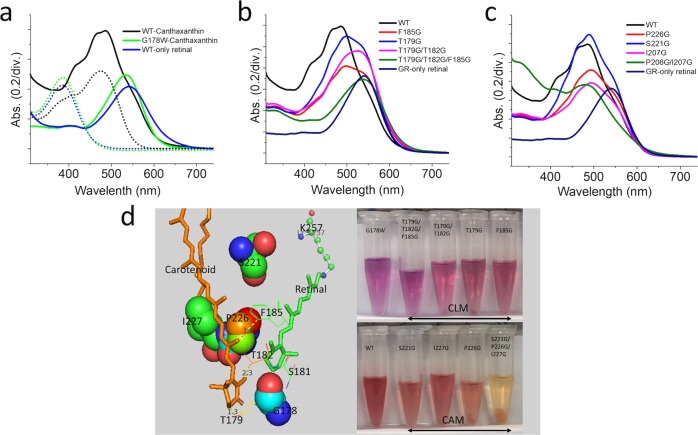
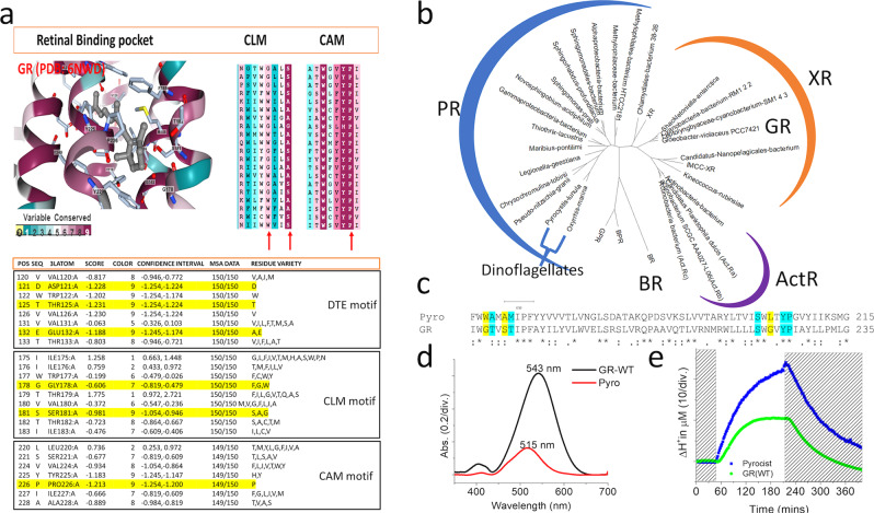
The position of carotenoid in xanthorhodopsin has been elucidated. However, a challenging expression of this opsin and a complex biosynthesis carotenoid in the laboratory hold back the insightful study of this rhodopsin. Here, we demonstrated co-expression of the xanthorhodopsin type isolated from Gloeobacter violaceus PCC 7421-Gloeobacter rhodopsin (GR) with a biosynthesized keto-carotenoid (canthaxanthin) targeting the carotenoid binding site. Direct mutation-induced changes in carotenoid-rhodopsin interaction revealed three crucial features: (1) carotenoid locked motif (CLM), (2) carotenoid aligned motif (CAM), and color tuning serines (CTS). Our single mutation results at 178 position (G178W) confirmed inhibition of carotenoid binding; however, the mutants showed better stability and proton pumping, which was also observed in the case of carotenoid binding characteristics. These effects demonstrated an adaptation of microbial rhodopsin that diverges from carotenoid harboring, along with expression in the dinoflagellate Pyrocystis lunula rhodopsin and the evolutionary substitution model. The study highlights a critical position of the carotenoid binding site, which significantly allows another protein engineering approach in the microbial rhodopsin family.


Conflict of interest statement

The authors declare no competing interests.

Figures

Fig. 1
Fig. 1. Co-expression of GR wild-type and mutants with canthaxanthin.
a GR wild-type with only retinal (blue line), GR-expressed with both retinal and carotenoid (black line), single mutation G178W expressed with both retinal and carotenoid (green line). Complete chromophore extraction from purified samples showed blue, black, and green (dot line), respectively. b Absorption spectra of the mutant on helix E near glycine 178, carotenoid locked motif. c Absorption spectra of mutant on helix F, carotenoid aligned motif. d Local view of targeting mutation site, a structure alignment of GR (pdb:6nwd) and XR (pdb:3ddl), and the purified GR variants co-expressed with retinal/carotenoid.
Fig. 2
Fig. 2. Structural-based alteration of targeting mutants and the interaction of the retinal and carotenoid chromophores.
a Maximum absorption shifted of targeted mutants. b The local view of mutation position shows in the red sphere Ser 221 for the strongest red-shifted and the blue sphere for Ser 181 for the strongest blue-shifted. c UV–vis spectra of wild type, S181G, and S221G. d Purified samples in 0.02% DDM. e Comparison of proton pumping with and without carotenoid, helix E mutants showed a reduction of proton pumping enhancement as a response to lower binding ration; however, helix F mutants with higher carotenoid binding ratio disrupted the benefit of carotenoid in proton pumping, suggesting the challenge of energy transfer and light absorption.
Fig. 3
Fig. 3. A single mutation G178W replaces and mimics the carotenoid binding.
a Comparison of proton pumping of GR wild-type, G187W mutant, and GR-carotenoid. b Retinal bleaching from opsin when exposed to high pH condition, G178W showed pH tolerance over GR-bound carotenoid and wilt-type. c Local view of G178W mutant and the polar interactive change of surrounding residues, while replacement of Gly to Trp make the overlap ring to the carotenoid 4-keto ring of carotenoid.
Fig. 4
Fig. 4. Divergent evolution of carotenoid binding in microbial rhodopsin.
a Multiple sequences alignment (MSA) calculated by Bayesian method for evolutionary substitution model of GR (pdb:6nwd) among 150 homologs. DTE motif is almost wholly conserved, while CLM motif showed Gly178 is replaced either bulky residue Trp or aromatic ring residue Phe, and at the same time Ser 181 is either replaced with tiny residue Gly or Ala, CAM motif showed completely conserved of retinal binding pocket Trp 222, and Pro 226, which suggests that this non-polar helix kink residue is essential. b The phylogenetic tree xanthorhodopsin, actinorhodopsin, bacteriorhodopsin, and proteorhodopsin, including dinoflagellate proteorhodopsin. XR showed evolutional closer distance to actinorhodopsin compared to proteorhodopsin. c One-to-one sequence alignment of dinoflagellate Pyrocis lununa rhodopsin and GR, Gly178 was replaced by Trp, and polar residue T179 and Ser181 were replaced by non-polar small alanine. d UV–vis spectra of purified Pyrocis lununa rhodopsin and GR, Pyro-rhodopsin showed maximum absorption at 515 nm in the more blue-green region compared to GR 543 nm. e The proton pumping of expressed GR and Pyro-rhodopsin in E. coli cells with the same protein concentration was calculated.

References

    1. Gómez-Consarnau L, et al. Microbial rhodopsins are major contributors to the solar energy captured in the sea. Sci. Adv. 2019;5:eaaw8855. doi: 10.1126/sciadv.aaw8855. - DOI - PMC - PubMed
    1. Govorunova EG, Sineshchekov OA, Li H, Spudich JL. Microbial rhodopsins: diversity, mechanisms, and optogenetic applications. Annu. Rev. Biochem. 2017;86:845–872. doi: 10.1146/annurev-biochem-101910-144233. - DOI - PMC - PubMed
    1. Ernst OP, et al. Microbial and animal rhodopsins: structures, functions, and molecular mechanisms. Chem. Rev. 2014;114:126–163. doi: 10.1021/cr4003769. - DOI - PMC - PubMed
    1. Oesterhelt D, Stoeckenius W. Rhodopsin-like protein from the purple membrane of Halobacterium halobium. Nat. N. Biol. 1971;233:149–152. doi: 10.1038/newbio233149a0. - DOI - PubMed
    1. Oesterhelt D, Stoeckenius W. Functions of a new photoreceptor membrane. Proc. Natl Acad. Sci. USA. 1973;70:2853–2857. doi: 10.1073/pnas.70.10.2853. - DOI - PMC - PubMed

Publication types