. 2022 Jun;24(6):845-857.
doi: 10.1038/s41556-022-00916-w. Epub 2022 May 30.

Polycomb repressive complex 2 shields naïve human pluripotent cells from trophectoderm differentiation

Affiliations

Polycomb repressive complex 2 shields naïve human pluripotent cells from trophectoderm differentiation

Banushree Kumar et al. Nat Cell Biol. 2022 Jun.

Abstract

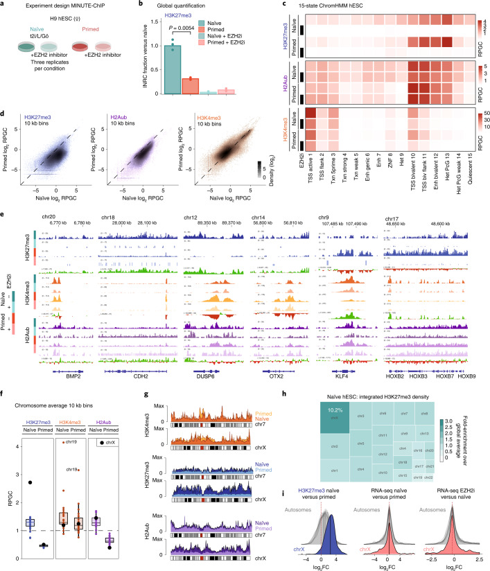
The first lineage choice in human embryo development separates trophectoderm from the inner cell mass. Naïve human embryonic stem cells are derived from the inner cell mass and offer possibilities to explore how lineage integrity is maintained. Here, we discover that polycomb repressive complex 2 (PRC2) maintains naïve pluripotency and restricts differentiation to trophectoderm and mesoderm lineages. Through quantitative epigenome profiling, we found that a broad gain of histone H3 lysine 27 trimethylation (H3K27me3) is a distinct feature of naïve pluripotency. We define shared and naïve-specific bivalent promoters featuring PRC2-mediated H3K27me3 concomitant with H3K4me3. Naïve bivalency maintains key trophectoderm and mesoderm transcription factors in a transcriptionally poised state. Inhibition of PRC2 forces naïve human embryonic stem cells into an 'activated' state, characterized by co-expression of pluripotency and lineage-specific transcription factors, followed by differentiation into either trophectoderm or mesoderm lineages. In summary, PRC2-mediated repression provides a highly adaptive mechanism to restrict lineage potential during early human development.


Conflict of interest statement

The authors declare no competing interests.

Figures

Fig. 1
Fig. 1. Diffuse H3K27me3 and H2Aub cover the naïve pluripotent genome.
a, Experimental design of MINUTE-ChIP experiment comparing hESCs in naïve and primed, untreated and treated (10 µM EZH2i) culture conditions. Three biological replicates of each condition were barcoded and combined into a single MINUTE-ChIP pool. See Extended Data Fig. 1a for a scheme of the MINUTE-ChIP workflow. b, Global levels of H3K27me3 as determined by MINUTE-ChIP input-normalized read counts (INRCs) in naïve or primed hESCs, cultured with or without EZH2 inhibitor. P values of pairwise comparisons (two-sided unpaired Student’s t-test) are given. See Extended Data Fig. 1b for corresponding analysis of H2Aub and H3K4me3. c, Histone H3K27me3, H3K4me3 and H2Aub levels by chromatin state (reads per genome coverage, RPGC). See Extended Data Fig. 1c for individual replicates. d, Genome-wide analysis of H3K27me3, H2Aub and H3K4me3 levels by 10 kb bins, comparing naïve and primed hESCs. e, Genome browser examples of genomic regions with differential occupancy of H3K27me3, H3K4me3 and/or H2Aub. Group-scaled histone modification signals and gain/loss tracks comparing naïve and primed signals are shown. f, Chromosome average enrichment of H3K4me3, H3K27me3 and H2Aub in naïve and primed hESCs. Box plot boxes show the 25th and 75th percentile with the median, and whiskers indicate 1.5 times the interquartile range. All individual data points are shown. g, Chromosome density plot comparing the X chromosome signals in naïve and primed hESCs to an autosome with similar size (chr7). See Extended Data Fig. 4c for a genome browser example of X chromosome region. h, Treemap showing a proportional representation of the total (integrated) amount of H3K27me3 by chromosome (area) and average density (colour intensity). i, Density plots of log2FC in promoter H3K27me3 levels (left) and RNA-seq output (middle) of genes grouped by chromosome, comparing naïve and primed hESCs. Density plots of log2FC in RNA-seq output of genes grouped by chromosome, comparing untreated and EZH2i-treated naïve hESCs (right). Median values by chromosome are given and indicated as vertical lines in the density plot. In cg, three combined biological replicates are shown. Source data
Fig. 2
Fig. 2. H3K27me3 is adaptive to gene expression changes between naïve and primed pluripotent states and contributes to repression of non-state-specific genes.
a, De novo annotation of bivalent promoters based on DESeq2 analysis. Five promoter classes were defined (for criteria, see Extended Data Fig. 5a): primed bivalent (Pr » Ni), naïve bivalent (Ni » Pr), common bivalent, H3K4me3 only and H3K4me3 negative (not shown). Average H3K27me3, H2Aub and H3K4me3 profile plots (fragments per genome coverage, FPGC) in naïve and primed hESCs for each class and corresponding heat maps for the first three states are shown. Profiles for EZH2i-treated naïve and primed conditions are shown as dashed lines. b, Alluvial plot showing H3K27me3 and H3K4me3 gains and losses (DESeq2 adjusted P < 0.05, fold change >1.5 from three replicates) at bivalent promoters between naïve and primed hESCs. Select connections are annotated. c, Context-specific transcriptional response to global H3K27me3 depletion in different classes of bivalent genes. RNA-seq changes (DESeq2 log2FC from three replicates each) are plotted, comparing naïve and primed conditions as well as EZH2i treatment and the respective control (left, naïve only; middle, primed only; right, shared bivalent promoters). The distribution of fold changes of all genes is shown as violin plots, and class-specific genes are shown as jitter points. The class-specific group was compared with all genes using a two-sided unpaired Wilcoxon test and Cohen’s d. d, Density plot of fold changes of H2Aub levels following H3K27me3 depletion in hESCs. Only genes that were derepressed upon EZH2i treatment (DESeq2 adjusted P < 0.05, fold change >1.5 based on three replicates) were included in the analysis. Bivalent promoters (hence including promoters of the naïve-bivalent and shared class) are compared with H3K27me3-devoid promoters. For an analysis of individual classes, see Extended Data Fig. 5b. e, Heat map showing RNA-seq expression (log2-transformed TPM) of previously defined marker genes for naïve and, primed pluripotency in naïve and primed cultures (± EZH2i treatment), as well as the H3K4me3 and H3K27me3 levels (RPGC) at their respective promoter. RPGC from combined replicates are shown for H3K4me3 and H3K27me3, whereas the individual replicate TPM values are plotted for RNA-seq data. n.s., not significant. Source data
Fig. 3
Fig. 3. PRC2 inhibition derepresses a naïve-specific subset of bivalent genes.
a, Volcano plot showing differentially expressed genes (DESeq2 FDR <5%, |log2FC| >1) between 7-day EZH2i-treated naïve hESCs and untreated hESCs. Bivalent gene classes as shown in Fig. 2a are indicated in red (primed bivalent), blue (shared bivalent) and teal (naïve bivalent). See Extended Data Fig. 5d for corresponding analysis in primed hESCs. b, Fractions of significant (FDR <5%) transcriptional changes in response to EZH2 inhibitor treatment of naïve and primed hESCs, among all or bivalent promoter classes. c, Intersection of genes derepressed after EZH2i treatment in naïve and primed hESCs. Each Venn intersection is further annotated by the H3K27me3 promoter class. d, Strip chart showing expression of a comprehensive set of marker genes defined from human embryo single-cell data comparing naïve hESCs and naïve hESCs treated with EZH2i. Markers are grouped into pre-lineage, ICM, epiblast, primitive endoderm (PrE), trophectoderm (TE), cytotrophoblast (CTB), syncytiotrophoblast (STB), extravillous trophoblast (EVT), amnion, extra-embryonic mesoderm (exMes), advanced mesoderm (AdvMes), emerging mesoderm (EmMes), nascent mesoderm (NasMes), axial mesoderm (AxMes) and primitive streak (PriS). Significant differences (DESeq2 FDR <5% from triplicates) are highlighted in blue (downregulated) or orange (upregulated). Source data
Fig. 4
Fig. 4. Loss of H3K27me3 in naïve hESCs activates trophectoderm gene expression programmes.
a, Heat map showing expression (TPM) in naïve and primed hESCs (± EZH2i treatment), as well as H3K4me3, H3K27me3 and H2Aub promoter status of selected trophectoderm and placenta-specific genes. RPGC from combined replicates are used for H3K4me3 and H3K27me3, whereas the three individual replicate TPM values are plotted for RNA-seq data. GATA3 binding as determined by ChIP–seq peaks during trophectoderm differentiation is indicated. For genome browser examples, see Extended Data Fig. 6a,c. b, Immunofluorescence confocal microscopy images of naïve H9 hESC colonies without treatment or with EZH2i (EPZ-6438) or EEDi (EED226) treatment for 7 days, assessing H3K27me3 levels, expression of pluripotency marker NANOG and trophectoderm transcription factor GATA3. Merged images show NANOG in purple, H3K27me3 in red, GATA3 in green, and Hoechst in blue. Scale bars, 10 µm. Data shown represent three independent experiments for EZH2i and two independent experiments for EEDi. For analogous experiment in naïve HS975 hESCs, see Extended Data Fig. 7a. c,d, CellProfiler image analysis of the experiment described in b. Per-nucleus H3K27me3 and GATA3 immunofluorescence intensities of a total of 670 nuclei derived from one experiment (165 untreated, 235 EZH2i and 270 EEDi). Box plot boxes show the 25th and 75th percentile with the median, and whiskers indicate 1.5 times the interquartile range. P values are determined by two-sided unpaired t-test (H3K27me3 P < 2.2 × 10⁻¹⁶ (top), GATA3 P = 2.4 × 10⁻⁵ for EZH2i and P = 0.046 for EEDi treatment). The threshold for GATA3+ cells (dashed line) is defined as 1.5× mean of untreated cells. Scatter plots contrasting per-nucleus GATA3 with H3K27me3 or NANOG immunofluorescence intensities. e, Scheme of experiment for CRISPR/Cas9-mediated acute deletion of EED. f, Representative image of experiment described in e. Example shown represents two independent experiments. Scale bars, 20 µm. g, CellProfiler image quantification of the experiment described in e. Scatter plots contrasting per-nucleus GATA3 and H3K27me3 immunofluorescence intensities of untransfected cells, NT gRNA or EED-targeting gRNA (EED gRNA) transfected cells. In total, 4,312 nuclei from two independent experiments were analysed. For statistics by individual colonies, see Extended Data Fig. 7b. Source data
Fig. 5
Fig. 5. Single-cell transcriptomic profiling of EZH2i-treated naïve and primed cells.
a, UMAP projection reference annotation (TE: trophectoderm; Exe_Mes: extra-embryonic mesoderm). The colour of each data point represents the cell annotations retrieved for each publication. Light-grey-coloured data points represent cells in the multi-data integration that were not used to infer cell annotations. The shape of data points for the embryonic cells indicates the data source. b UMAP projection and cell identity annotations of single-cell transcriptome datasets obtained for EZH2i-treated naïve and primed cells at indicated timepoint, showing ELCs, TLCs, MeLCs, AMLCs and HLCs. Percentage of cells within major clusters are indicated in the maps. Bar chart (bottom) shows the percentage of cells per cluster at each time point. c, UMAP projection and cell identity annotations as in b for primed hESCs treated with EZH2i. In ac, UMAP was generated from an extensive reference dataset, including data from human embryonic and PASE model (Extended Data Fig. 8a,b and Methods). d, Top ten significantly upregulated genes in MeLCs or TLCs, compared with ELC population. Bubble plot shows proportion of expressing cells and average expression per gene and condition. e, Single-cell expression of select TLC and MeLC markers mapped onto UMAP. log2 expression of selected genes overlaid on the UMAP. f, Selected genes differentially expressed within ELC population comparing 7-day-EZH2i-treated samples and untreated samples. Bubble plot shows proportion of expressing cells and average expression per gene and condition. g, Single-cell log2 expression of select factors mapped onto UMAP. Source data
Fig. 6
Fig. 6. Trajectory inference and gene expression dynamics for EZH2i-treated naïve hESCs.
a,b, Trajectory inference colour-coded by inferred pseudotime and cell types. c, Further subclassification of ELC cells based on cell state and pseudotime distribution: gELC, aELC, TaELC and MaELC. Ridge plot (bottom) shows distribution of single cells along the pseudotime axis for naïve hESCs untreated or treated for 2, 4 and 7 days with EZH2i. Two dashed lines on ridge plot represent the pseudotime for transition from gELC to aELC, and bifurcation into MaELC and TaELC, respectively. d, Cells from untreated and EZH2i-treated (2, 4 and 7 days) naïve hESCs mapped onto trajectory. e, Volcano plot showing differentially expressed genes (24 up and 27 down) comparing aELC and gELC population (combined data from 4-day and 7-day EZH2i treatment of naïve hESCs). f, Volcano plot showing differentially expressed genes comparing mesoderm (MaELC, 67 genes) and trophectoderm (TaELC, 27 genes) branching at bifurcation point in 7-day-EZH2i treated naïve hESCs. g, Expression dynamics (pseudotime) of selected genes during EZH2i treatment on naïve cells. The confidence interval (95%) is indicated by bandwidth. MeLC branch and TLC branch are indicated by pink and blue lines, respectively. Source data
Extended Data Fig. 1
Extended Data Fig. 1. Scheme of MINUTE-ChIP workflow.
a Scheme of the MINUTE-ChIP workflow. Triplicate cell pellets for each condition were lysed and the chromatin was enzymatically fragmented to mono- and di- nucleosomes and barcoded by ligating on a dsDNA adapter. Samples were then pooled and the barcoded lysate was aliquoted to individual ChIP reactions (5% of the ChIP volume was reserved as input material and carried through protocol in a manner similar to the IPs) with magnetic beads pre-coupled with the respective antibodies. Subsequently, beads were washed and IPed DNA was eluted, proteinase K digested and purified. For constructing the final libraries, DNA from each IP was in vitro transcribed using the T7 promoter in the adapter that was ligated on in the initial step. The resulting RNA was appended with an RNA 3’ adapter (RA3) allowing for specific paired end sequencing. The RA3 in turn was used to prime the reverse transcription reaction, generating cDNA that was used as a template for the final low-cycle library PCR. At this stage, in addition to the Illumina-compatible sequences, PCR primers also carried a second barcode sequence to serve as an identifier for the IP performed. Finally, libraries are pooled and sequenced on the Illumina platform,. b Global genome-wide levels of H3K4me3 and H2Aub as determined by input-normalized total read counts (INRC) in naïve or primed hESC, cultured with or without EZH2 inhibitor. P values of pairwise comparisons (two-sided unpaired Student’s t test) are given. n = 3 biologically independent samples. Tracks for subsequent analysis are scaled according to the INRC values as reads per genome coverage (RPGC), with naïve serving as a reference scaled to 1x genome coverage (global average equals to 1 RPGC) c Histone H3K27me3, H3K4me3 and H2Aub levels by chromatin state. RPGC of individual replicates are shown. d Genome-wide correlation (10 kb bins, Persson-correlation coefficient) of MINUTE-ChIP replicates, including corresponding inputs. e Exemplary genome-wide comparison of 10 kb bins as scatter plot. Source data
Extended Data Fig. 2
Extended Data Fig. 2. H3K27me3 levels in naïve and primed hESC.
a Phase contrast microscopy images of primed and naïve hESC, untreated or treated with EZH2i over 5 days showing similar growth and morphology. Data shown represent 2 independent experiments. Scale bars 100 µm. b Immunofluorescence microscopy showing H3K27me3 staining of naïve and primed hESC + /- EZH2i treatment. Stainings were performed in parallel in the same antibody dilutions and images were acquired with the same gain settings to allow a quantitative comparison. Data shown represent 2 independent experiments. Scale bars 10 µm. c Representative western blot for quantification of H3K4me3, H2Aub and H3K27me3 assayed using two-color IR western blot, in hESC and WT(J1) mouse ESC (grown in 2i/Serum) with and without EZH2i treatment. d Quantification of western blots, represented as mean of two biological replicates. Representative image shown. We note that the discrepancy in fold-change comparing naïve and primed hESC with different quantitative methodologies MINUTE-ChIP, IF, western blots may arise from method-specific threshold sensitivity, dose-response curves and signal saturation levels. MINUTE-ChIP measures H3K27me3-density on the level of nucleosomes, whereas western blot yields a per-histone level quantification. We have previously confirmed that MINUTE-ChIP signal is linearly proportional under the dynamic range relevant to the H3K27me3 levels assayed. e Time course of H3K27me3-depletion using immunofluorescence microscopy. Boxplot showing quantification performed using CellProfiler image analysis of 1172 nuclei (237 untreated, 140 day 2, 314 day 4, 451 day 7) derived from 1 experiment. Box plot boxes show the 25th and 75th percentile with the median, and whiskers indicate 1.5x the interquartile range. P values determined by two-sided unpaired t test (respectively p = 1.2e-7, p < 2.22e-16, p < 2.22e-16). f Representative immunofluorescence microscopy image for e), scale bar 10 µm. Source data
Extended Data Fig. 3
Extended Data Fig. 3. Genome-wide histone posttranslational modification changes between naïve and primed hESC.
a Fraction of genome-wide 10 kb windows that significantly (DESeq2 p.adj < 0.05 and fold-change > 1.5 from three replicates) gain or lose H3K27me3, H2Aub or H3K4me3 levels between naïve and primed state. b Fraction of promoters (all or bivalent) that significantly (DESeq2 p.adj < 0.05 and fold-change > 1.5 from three replicates) gain or lose H3K27me3, H2Aub or H3K4me3 levels between naïve and primed state. c Scatter plots showing H3K27me3, H2Aub or H3K4me3 levels (log2-transformed RPGC) at promoters in naïve versus primed hESC. Bivalent genes are highlighted in black. d Scatter plots showing the relation of H3K27me3 and H2Aub across genome-wide 10 kb bins in naïve and primed hESC, as well as the correlation of changes. 10 kb bins overlapping with bivalent TSS are highlighted in turquoise e Scatter plot showing the relation of H2Aub across genome-wide 10 kb bins in naïve vs. EZH2i-treated naïve hESC. Significant bins are highlighted in red. 10 kb bins overlapping with bivalent promoters are highlighted in turquoise. Fraction of 10 kb bins significantly changing (DESeq2 p.adj < 0.05 and fold-change > 1.5 from three replicates) are given for all 10 kb bins, 10 kb bins overlapping with promoters, and 10 kb bins overlapping with bivalent promoters. Scatter plot showing the relation of H2Aub across promoters in naïve vs. EZH2i-treated naïve hESC. Significant bins are highlighted in red. Bivalent promoters are highlighted in black. Source data
Extended Data Fig. 4
Extended Data Fig. 4. High H3K27me3 density on naïve X chromosomes.
a Representative Genome browser overview showing H3K27me3 density across all chromosomes. Published H3K27me3 ChIP and MINUTE-ChIP H3K27me3 tracks (combined replicates), as well as MINUTE-ChIP input tracks are shown. b chromosome average enrichment of H3K4me3, H3K27me3 and H2Aub in naïve and primed hESC, cultured with or without EZH2i for 7 days. RPGC of combined replicates is shown. Box plot boxes show the 25th and 75th percentile with the median, and whiskers indicate 1.5x the interquartile range. c Genome browser view of the XIST neighborhood on the X chromosome at megabase resolution. Shown is the combined signal of three replicates for H3K27me3, H3K4me3, H2Aub as well as gain/loss tracks comparing naïve and primed signal, and stranded RNA-Seq signal (one of three replicates). Tracks from the same histone modification are shown on the same RPGC scale. RNA-Seq expression is shown on the same scale. Strong H3K27me3 accumulation in large blocks on naïve X chromosome is evident, with the exception of some islands that typically harbor genes active in naïve state, such as the XIST locus, which do not accumulate H3K27me3. d MA-plot of RNA-Seq data (Base mean and log2 fold-change as calculated with DESeq2 from triplicates) comparing naïve and primed hESC. Genes on the X chromosome are highlighted in black with selected annotations. e MA-plot of RNA-Seq data (Base mean and log2 fold-change as calculated with DESeq2 from triplicates) comparing untreated and EZH2i-treated naïve hESC. Genes on the X chromosome are highlighted in black (padj < 0.05 highlighted in red) with selected annotations. f H3K27me3 levels at promoters on the X chromosome or autosomes in naïve hESC + /- EZH2i treatment (top). Two groups of promoters are plotted: those of genes upregulated with EZH2i treatment in naïve hESC and those downregulated. Expression levels (log2-transformed TPM) at genes on the X chromosome or autosomes in naïve and primed hESC + /- EZH2i treatment (bottom). Box plot boxes show the 25th and 75th percentile with the median, and whiskers indicate 1.5x the interquartile range. Source data
Extended Data Fig. 5
Extended Data Fig. 5. Defining shared and state-specific bivalent promoters in naïve and primed hESC.
a Differential occupancy H3K27me3 at promoters using DESeq2. Volcano plot (DESeq2 based on three replicates) comparing promoter H3K27me3 levels between naïve and primed hESC. Explanation of criteria for defining naïve-bivalent, primed-bivalent and common bivalent gene classes. b Density plot of fold-changes of H2Aub levels following H3K27me3 depletion in hESC. Only genes that were derepressed upon EZH2i-treatment (DESeq2 p.adj < 0.05, fold-change > 2 based on three replicates) were included in the analysis. The different classes of bivalent promoters as defined above are compared to H3K27me3-devoid promoters. c Genome browser view of TFAP2A and TFAP2C transcription factors in naïve and primed hESC + /− 7d EZH2i treatment. Shown is the combined signal of three replicates for H3K27me3, H3K4me3, H2Aub as well as gain/loss tracks comparing naïve and primed signal, and stranded RNA-Seq signal (one of three replicates shown). Tracks from the same histone modification are shown on the same RPGC scale. RNA-Seq expression is shown on the same TPM scale. d Volcano plot with selected annotations showing differentially expressed genes (DESeq2 FDR < 5%, |log2FC | > 1) between 7d EZH2i-treated and untreated primed hESC. Bivalent gene classes are shown as colored dots: red (primed-bivalent), blue (shared-bivalent), teal (naïve bivalent).e Stripchart showing expression of a comprehensive set of marker genes defined from human embryo single cell data comparing primed hESC + /− 7d EZH2i. Markers are grouped into pre-lineage, inner cell mass (ICM), epiblast, primitive endoderm, (PrE), trophectoderm (TE) cytotrophoblast (CTB), syncytiotrophoblast (STB), extravillous trophoblast (EVT), amnion, extraembryonic mesoderm (exMes), advanced mesoderm (AdvMes), emerging mesoderm (EmMes), nascent mesoderm (NasMes), axial mesoderm (AxMes) and primitive streak (PriS). Significant differences (DESeq2 p.adj < 0.05 from triplicates) are highlighted in color. f Immunofluorescence confocal microscopy images of primed H9 hESC colonies with or without EZH2i (EPZ-6438) treatment for 7 days, stained for pluripotency markers NANOG, SOX2 and OCT3/4 (right panel) and H3K27me3 and trophectoderm transcription factor GATA3 (left panel). Data shown represent 2 independent experiments. Scale bars 50 µm. Source data
Extended Data Fig. 6
Extended Data Fig. 6. Loss of H3K27me3 in naïve hESC activates trophectoderm gene expression programs.
a Genome browser examples of selected bivalent trophectoderm lineage markers. Shown is the combined signal of three replicates for H3K27me3, H3K4me3, H2Aub, RNA-Seq, as well as gain/loss tracks comparing naïve and primed signals. Tracks from the same histone modification are shown on the same RPGC scale. RNA-Seq expression from one replicate is shown on the same TPM scale. b Venn diagram intersecting gene sets: 1894 significantly upregulated genes in naïve hESC treated with EZH2i, 758 significantly downregulated genes in naïve hESC treated with EZH2i, 1831 genes with promoter-proximal GATA3 binding sites. c Genome browser examples of selected trophectoderm genes with GATA3 binding sites. Shown is the combined signal of three replicates for H3K27me3, H3K4me3, H2Aub, RNA-Seq, as well as gain/loss tracks comparing naïve and primed signals. Tracks from the same histone modification are shown on the same RPGC scale. RNA-Seq expression from one replicate is shown on the same TPM scale.
Extended Data Fig. 7
Extended Data Fig. 7. GATA3 induction upon pharmacological and genetic targeting of PRC2.
a Immunofluorescence microscopy (on Olympus IX81, 20x magnification) showing H3K27me3 and GATA3 staining of naïve HS975 hESC cultured in 5iLAF medium with or without EZH2i (EPZ-6438) treatment for 7 days. Scale bars are 50 µm. Tukey boxplot generated from corresponding CellProfiler image analysis shows per-nucleus H3K27me3 and GATA3 intensities over n = 195 nuclei (69 untreated, 127 EZH2i) derived from 1 experiment. EZH2i treatment resulted in 6.3% GATA3 + nuclei for this cell type. The threshold for GATA3 + cells (dashed line) is defined as 1.5x mean of untreated cells. Box plot boxes show the 25th and 75th percentile with the median, and whiskers indicate 1.5x the interquartile range. P values estimated by two-sided unpaired t test (p = 0.047; p < 2.2e-16, respectively). b Boxplots show per-nucleus EED (top panel) and GATA3 (bottom panel) intensities summarized by the imaged colonies for CRISPR/Cas9 based acute targeting of EED. corresponding to Fig. 4f. Box plot boxes show the 25th and 75th percentile with the median, and whiskers indicate 1.5x the interquartile range. c Immunofluorescence microscopy showing H3K27me3 and GATA3 staining of naïve H9 hESC cultured in t2iLGö (NaiveCult) or PXGL medium with or without EZH2i (EPZ-6438) treatment for 7 days. Scale bar is 20 µm. Tukey boxplot generated from corresponding CellProfiler image analysis shows per-nucleus H3K27me3 and GATA3 intensities over 3045 nuclei in total (500 t2iLGö, 846 t2iLGö +EZH2i, 1037 PXGL, 662 PXGL + EZH2i) derived from 1 experiment. The threshold for GATA3 + cells (dashed line) is defined as 1.5x mean of untreated cells. Box plot boxes show the 25th and 75th percentile with the median, and whiskers indicate 1.5x the interquartile range. Source data
Extended Data Fig. 8
Extended Data Fig. 8. scRNA-seq UMAP cluster assignment and characterization.
a UMAP projection of integrated datasets as in Fig. 5a, highlighting cells from Zheng et al. b Unassigned Seurat cluster distribution of UMAP used in Fig. 5a. c Bubble plot showing expression of key marker genes across the cell clusters as indicated. The sizes and colors of dots indicate the proportion of cells expressing the corresponding genes and their averaged scaled values of log-transformed expression, respectively. d Reanalysis of published scRNA-seq in naïve hESC from Messmer et. al. Expression of GATA3, CDX1, HAND1, KRT19 is color coded in PCA map as defined in Messmer et. al. e Volcano plot showing differentially expressed genes (482 up, 387 down) comparing ELC population at 7d EZH2i with ELC population in untreated naïve hESC. f Violin plot showing single-cell log-transformed expression of HAND1 and CDX1 transcription factors. Source data
Extended Data Fig. 9
Extended Data Fig. 9. Epigenomic landscape of differentially expressed genes in ELC population after 7d EZH2i treatment.
a Heatmap showing RNA-Seq expression levels (log2-transformed TPM) of core pluripotency markers in naïve and primed hESC + /− 7d EZH2i treatment, as well as the H3K4me3 and H3K27me3 levels (RPGC) at their respective promoter. RPGC from combined replicates are used for H3K4me3 and H3K27me3, whereas the three individual replicate TPM values are plotted for RNA-Seq data. b Genome browser view of TBXT, HAND1 and CDX1 transcription factors in naïve and primed hESC + /− 7d EZH2i treatment. Shown is the combined signal of three replicates for H3K27me3, H3K4me3, H2Aub as well as gain/loss tracks comparing naïve and primed signal, and stranded RNA-Seq signal. Tracks from the same histone modification are shown on the same RPGC scale. RNA-Seq expression is shown on the same TPM scale. Published GATA3 ChIP-seq tracks are shown. c Trajectory inference colored-coded by inferred cell state calculated by monocle. d Indicated cell populations as defined in Fig. 5b are mapped onto the trajectory (in red). Source data

Comment in

References

    1. Thomson, J. A. et al. Embryonic stem cell lines derived from human blastocysts. Science282, 1145–1147 (1998). - PubMed
    1. Xu, R.-H. et al. BMP4 initiates human embryonic stem cell differentiation to trophoblast. Nat. Biotechnol.20, 1261–1264 (2002). - PubMed
    1. Das, P. et al. Effects of fgf2 and oxygen in the bmp4-driven differentiation of trophoblast from human embryonic stem cells. Stem Cell Res.1, 61–74 (2007). - PMC - PubMed
    1. Marchand, M. et al. Transcriptomic signature of trophoblast differentiation in a human embryonic stem cell model. Biol. Reprod.84, 1258–1271 (2011). - PubMed
    1. Bai, Q. et al. Dissecting the first transcriptional divergence during human embryonic development. Stem Cell Rev.8, 150–162 (2012). - PMC - PubMed

Publication types

MeSH terms

Grants and funding