. 2022 May 30;19(1):37.
doi: 10.1186/s12987-022-00334-y.

Polarized α-synuclein trafficking and transcytosis across brain endothelial cells via Rab7-decorated carriers

Affiliations

Polarized α-synuclein trafficking and transcytosis across brain endothelial cells via Rab7-decorated carriers

Parvez Alam et al. Fluids Barriers CNS. .

Abstract

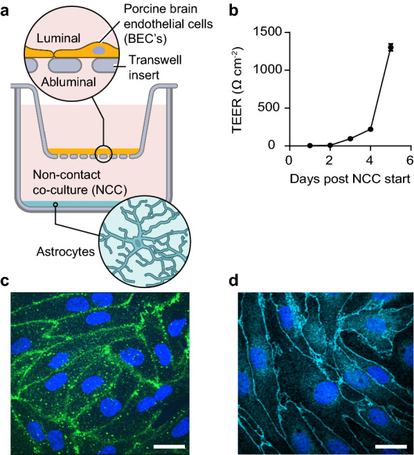
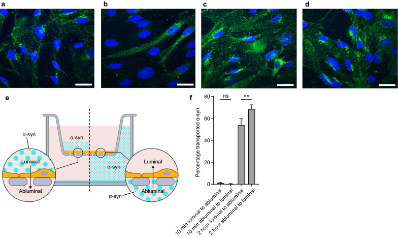
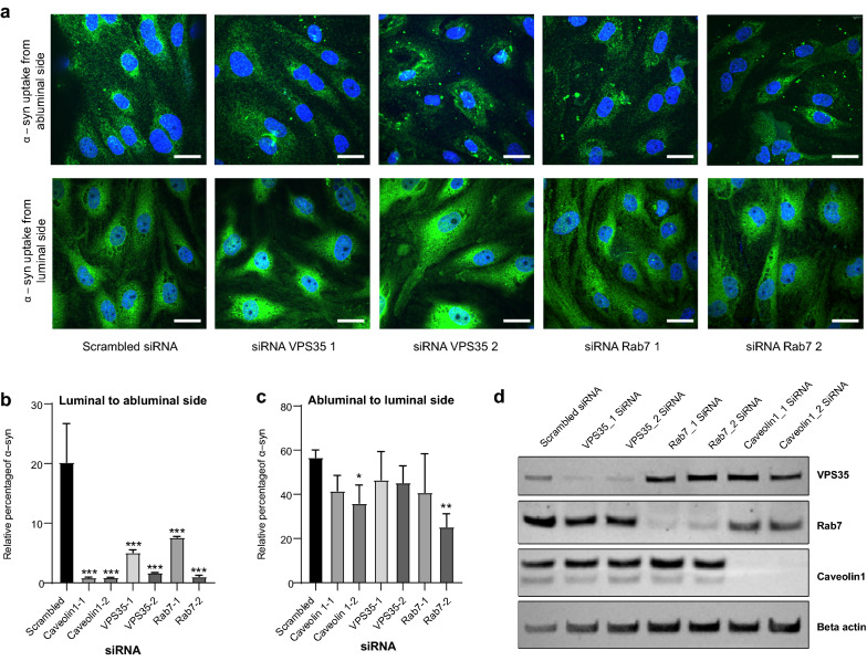
Parkinson's disease is mainly caused by aggregation of α-synuclein (α-syn) in the brain. Exchange of α-syn between the brain and peripheral tissues could have important pathophysiological and therapeutic implications, but the trafficking mechanism of α-syn across the blood brain-barrier (BBB) remains unclear. In this study, we therefore investigated uptake and transport mechanisms of α-syn monomers and oligomers across an in vitro BBB model system. Both α-syn monomers and oligomers were internalized by primary brain endothelial cells, with increased restriction of oligomeric over monomeric transport. To enlighten the trafficking route of monomeric α-syn in brain endothelial cells, we investigated co-localization of α-syn and intracellular markers of vesicular transport. Here, we observed the highest colocalization with clathrin, Rab7 and VPS35, suggesting a clathrin-dependent internalization, preferentially followed by a late endosome retromer-connected trafficking pathway. Furthermore, STED microscopy revealed monomeric α-syn trafficking via Rab7-decorated carriers. Knockdown of Caveolin1, VPS35, and Rab7 using siRNA did not affect monomeric α-syn uptake into endothelial cells. However, it significantly reduced transcytosis of monomeric α-syn in the luminal-abluminal direction, suggesting a polarized regulation of monomeric α-syn vesicular transport. Our findings suggest a direct role for Rab7 in polarized trafficking of monomeric α-syn across BBB endothelium, and the potential of Rab7 directed trafficking to constitute a target pathway for new therapeutic strategies against Parkinson's disease and related synucleinopathies.

Keywords: Brain endothelial cells and blood brain barrier; Parkinson’s disease; Polarization trap; Rab7; Retromer; Transcytosis; α-synuclein.


Conflict of interest statement

The authors declare no competing interests.

Figures

Fig. 1
Fig. 1
The experimental BBB model. Schematic for the in vitro BBB model of pBECs (porcine brain endothelial cells) in Non-Contact Co-culture (NCC) with astrocytes (a). Validation of the model with representative TEER (trans-endothelial cells resistance) values post NCC days (b). Representative micrographs of immunostainings for tight junction marker proteins showing (c) Claudin 5 (c) and ZO-1 (d). Scale bars show 15 µm
Fig. 2
Fig. 2
Transport of α-syn monomers through the BBB model. Representative confocal micrographs of α-syn monomer added to pBECs seeded on filters. Green are stains for the α-syn monomer and blue are Hoechst stains to mark the nucleus of the cells. a 10 min luminal side (b) 10 min abluminal side (c) 2 h luminal side and (d) 2 h abluminal side, scale bars show 15 µm. e Experimental set up: 100 nM α-syn monomer was added on to the BBB cell model and incubated for 10 min or 2 h at 37 °C with shaking at 150 rpm (f) ELISA results for α-syn transport across BBB when added from the luminal or abluminal side. Significance was tested using One-way ANOVA with Šidák´s multiple comparisons test
Fig. 3
Fig. 3
Transport of α-syn oligomers through the BBB model. Representative confocal microscopy images of α-syn ONE oligomers uptake in pBEC on filters after 2 h chase. Green color are stains for α-syn oligomers and blue are Hoechst stains. a α-syn oligomer added from luminal side (b) α-syn oligomer added from abluminal side (c). Scale bars show 15 µm. ELISA results for α-syn oligomeric transport across BBB model with 2 h of chase for the indicated directions. Significance was tested using One-way ANOVA with Šidák´s multiple comparisons test
Fig. 4
Fig. 4
Analysis of α-syn co-occurrence with trafficking markers. Representative confocal micrographs of α-syn monomer treated pBECs on filters with α-syn added for 10 min to the luminal side (a). Green show α-syn monomer stain, blue is Hoechst stain and red is the EEA1 stain. Lower right micrograph shows the outcome of IMARIS spot segmentation and colocalization analysis between α-syn and EEA1 channels. Scale bars show 15 µm. b Semi-quantification of α-syn co-occurrence with different compartments in pBEC: EEA1 (early endosome), RAB7 (endosome-lysosome), VPS35 (endosome-golgi, retromer), Rab8a (Golgi-plasma membrane), Caveolin1 (Caveolae/transendothelial channels) and Clathrin (endo-lysosomal vesicles). Statistical difference was tested using an ordinary two-way ANOVA followed by Tukey's multiple comparisons test
Fig. 5
Fig. 5
Effect of intracellular trafficking machinery on α-syn transport through the BBB model. Representative micrographs of α-syn uptake in pBEC’s on filters knocked down for VPS35 and Rab7 and treated for 2 h with monomeric α -syn, as indicated in (a). Scale bars show 15 µm. Relative percentage of α-syn transcytosis in cells pretreated with indicated siRNA (in comparison to scrambled siRNA) and treated for 2 h with α-syn from luminal (b) and abluminal (c) side of the BBB model, respectively. Bar plots in (b, c) show mean values of ELISA measurements. Statistical difference was tested using an ordinary one-way ANOVA followed by Dunnett's multiple comparisons test. Representative Western blot to confirm the efficiency of the siRNA mediated knockdown of indicated proteins in pBECs on Transwell filters
Fig. 6
Fig. 6
Rab7 decorates α-syn carriers. Quantification of Rab7 and VPS35 co-localization with ɑ-syn added from the luminal or abluminal side. a, b are micrographs of pBEC cells with luminally and abluminally added ɑ-syn, respectively, co-stained for Rab7. d, e are micrographs of pBEC cells with luminally and abluminally added ɑ-syn, respectively, co-stained for VPS35. Representative dual-color confocal micrograph (i) with Rab7 or VPS35 (red) and ɑ-syn (green), scale bar show 5 µm. Maximum z-projected 3D STED stack (ii) of the highlighted area in (i), scale bar show 1 µm. Micrographs (zx-i) and (zx-ii) are sideview representations of the cells from maximum y-projections of the 400 nm slices highlighted in (ii). Scale bar show 1 µm. All images are contrast adjusted to enhance clarity. Pearson’s correlation coefficients between Rab7 and ɑ-syn are shown in (c) and between VPS35 and ɑ-syn in (f)

Similar articles

Cited by

References

    1. Dauer W, Przedborski S. Parkinson's disease: mechanisms and models. Neuron. 2003;39(6):889–909. doi: 10.1016/S0896-6273(03)00568-3. - DOI - PubMed
    1. McCann H, Stevens CH, Cartwright H, Halliday GM. α-Synucleinopathy phenotypes. Parkinsonism Relat Disord. 2014;20:S62–S67. doi: 10.1016/S1353-8020(13)70017-8. - DOI - PubMed
    1. Spillantini MG, Crowther RA, Jakes R, Hasegawa M, Goedert M. α-Synuclein in filamentous inclusions of Lewy bodies from Parkinson’s disease and dementia with Lewy bodies. Proc Natl Acad Sci. 1998;95(11):6469–6473. doi: 10.1073/pnas.95.11.6469. - DOI - PMC - PubMed
    1. Bisaglia M, Mammi S, Bubacco L. Structural insights on physiological functions and pathological effects of±-synuclein. FASEB J. 2009;23(2):329–340. doi: 10.1096/fj.08-119784. - DOI - PubMed
    1. Alam P, Bousset L, Melki R, Otzen DE. α-synuclein oligomers and fibrils: a spectrum of species, a spectrum of toxicities. J Neurochem. 2019;150(5):522–534. doi: 10.1111/jnc.14808. - DOI - PubMed