. 2022 May 30;26(1):155.
doi: 10.1186/s13054-022-04002-3.

Impairment of neutrophil functions and homeostasis in COVID-19 patients: association with disease severity

Affiliations

Impairment of neutrophil functions and homeostasis in COVID-19 patients: association with disease severity

Chloé Loyer et al. Crit Care. .

Abstract

Background: A dysregulated immune response is emerging as a key feature of critical illness in COVID-19. Neutrophils are key components of early innate immunity that, if not tightly regulated, contribute to uncontrolled systemic inflammation. We sought to decipher the role of neutrophil phenotypes, functions, and homeostasis in COVID-19 disease severity and outcome.

Methods: By using flow cytometry, this longitudinal study compares peripheral whole-blood neutrophils from 90 COVID-19 ICU patients with those of 22 SARS-CoV-2-negative patients hospitalized for severe community-acquired pneumonia (CAP) and 38 healthy controls. We also assessed correlations between these phenotypic and functional indicators and markers of endothelial damage as well as disease severity.

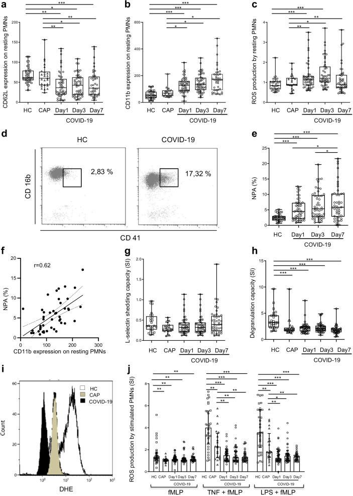
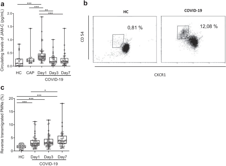
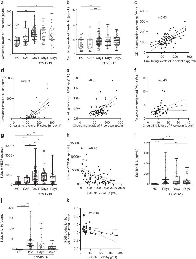
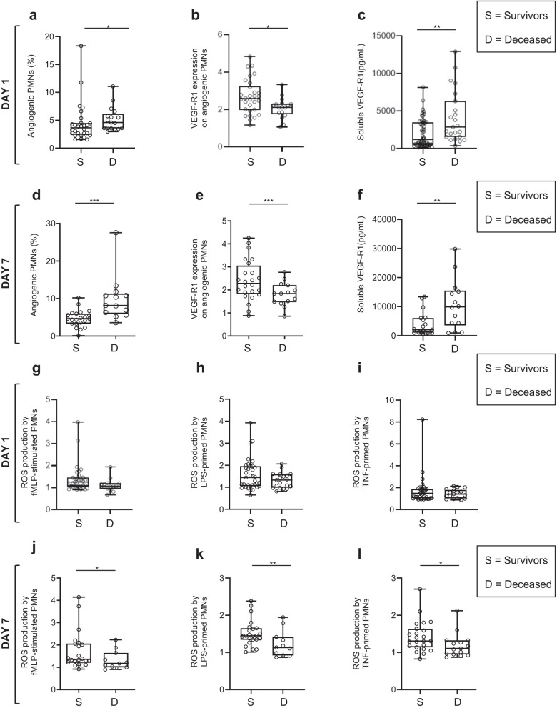
Results: At ICU admission, the circulating neutrophils of the COVID-19 patients showed continuous basal hyperactivation not seen in CAP patients, associated with higher circulating levels of soluble E- and P-selectin, which reflect platelet and endothelial activation. Furthermore, COVID-19 patients had expanded aged-angiogenic and reverse transmigrated neutrophil subsets-both involved in endothelial dysfunction and vascular inflammation. Simultaneously, COVID-19 patients had significantly lower levels of neutrophil oxidative burst in response to bacterial formyl peptide. Moreover patients dying of COVID-19 had significantly higher expansion of aged-angiogenic neutrophil subset and greater impairment of oxidative burst response than survivors.

Conclusions: These data suggest that neutrophil exhaustion may be involved in the pathogenesis of severe COVID-19 and identify angiogenic neutrophils as a potentially harmful subset involved in fatal outcome.

Keywords: Angiogenic neutrophils; COVID-19; Neutrophil; Oxidative burst; Vascular inflammation.


Conflict of interest statement

The authors declare that they have no competing interests.

Figures

Fig. 1
Fig. 1
Phenotypic and functional characterization of circulating neutrophils from COVID-19 patients. a, b Surface expression of CD62L (a) and CD11b (b) on resting neutrophils (PMNs) was studied in whole-blood samples maintained at 4 °C and stained with specific monoclonal antibodies. Results are expressed as mean fluorescence intensity (MFI). c Production of ROS by unstimulated neutrophils was studied with dihydroethidium (DHE) oxidation after treatment of whole-blood samples for 50 min with PBS; results are expressed as MFI. df Analysis of neutrophil-platelet aggregates (NPA). d Gating strategy and representative dot plots of flow cytometry analysis. e NPA levels, expressed as percentage of neutrophils that bind platelets. f Correlation of the percentage of NPA with CD11b expression at the neutrophil surface. g, h Analysis of capacity for L-selectin shedding (g) and neutrophil degranulation (h) in response to stimulation. CD62L and CD11b expression were analyzed after incubation of whole-blood samples for 45 min with PBS or fMLP (10−6 M). Results are expressed as a stimulation index (SI; MFI of stimulated sample/MFI of unstimulated sample). i, j ROS production by stimulated neutrophils was measured after pretreatment of whole-blood samples for 45 min with PBS, TNF-α (TNF, 5 ng/mL) or LPS (TLR4 agonist, 10 ng/mL). One histogram representative of ROS production by LPS-primed samples from a control (white), a CAP patient (grey) and a COVID-19 patient (black) (i). Results are expressed as SI (j). Samples came from age-matched healthy controls (HCs) (n = 38), CAP patients (n = 22) and COVID-19 patients at day 1 (n = 53), day 3 (n = 49) and day 7 (n = 40). Values are means ± SEM. *P < 0.05, **P < 0.01, ***P < 0.001, adjusted for age
Fig. 2
Fig. 2
Impaired homeostasis of circulating neutrophils in COVID-19 patients. ac Analysis of senescent and immunosuppressive neutrophil subsets in COVID-19 patients. Whole-blood samples were incubated for 45 min at 4 °C with Pe-Cy7-anti-human CXCR4, PE-anti-human CD11b, and APC-anti-human CD62L (a) or with FITC-anti-human CD16, PE-anti-human CD11c, Pe-Cy7-anti-human CD11b, and APC-anti-human CD62L (b) antibodies. a Percentages of the CXCR4bright/CD62Ldim senescent PMN subset. b Percentages of the CD16bright/CD62Ldim immunosuppressive PMN subset. (c) Ratio between the senescent and the immunosuppressive PMN subsets. dh Analysis of the angiogenic neutrophil subset in COVID-19 patients. Whole-blood samples were incubated for 45 min at 4 °C with FITC-anti-human VEGF-R1 and BV-481-anti-human CD49d. d Representative dot plot of angiogenic CD49dbright neutrophils gated according to forward-scattered light (FSC)/CD49d expression. e Percentages of the CD49dbright angiogenic neutrophil subset. f Expression of CD49d expression was analyzed at the surface of angiogenic neutrophils (CD49dbright); results are expressed in MFI. g One histogram representative of VEGF-R1 expression on angiogenic neutrophils from a control (white) and a COVID-19 patient (black). h Expression of VEGF-R1 on angiogenic neutrophils; results are expressed as MFI. Samples came from age-matched healthy controls (HCs) (n = 38), CAP patients (n = 22) and COVID-19 patients at day 1 (n = 53), day 3 (n = 49) and day 7 (n = 40). i Soluble VEGF-R1 was quantified by ELISA in HCs and COVID-19 patients at day 1 (n = 82), day 3 (n = 33) and day 7 (n = 32); results are as pg/ml. Values are means ± SEM. j Correlation between expression of VEGF-R1 on angiogenic neutrophils and soluble VEGF-R1 in COVID-19 patients. *P < 0.05, **P < 0.01, ***P < 0.001, adjusted for age. Samples came from age-matched healthy controls (HCs) (n = 38), CAP patients (n = 22) and COVID-19 patients at day 1 (n = 53), day 3 (n = 49) and day 7 (n = 40)
Fig. 3
Fig. 3
COVID-19 patients have higher levels of soluble JAM-C and of neutrophil reverse transendothelial transmigration. a Soluble JAM-C (sJAM-C) was quantified by ELISA; results are expressed as pg/ml. b, c Quantification of neutrophils undergoing reverse-endothelial transmigration (reverse transmigrated neutrophils, rTEM). Whole-blood samples were incubated for 45 min at 4 °C with FITC-anti-human CD181 (CXCR1) and PE-anti-human CD54 antibodies. b Representative dot plots of the neutrophil phenotype according to CXCR1 and CD54 expression in an HC (left) and a COVID-19 patient (right); c Percentage of rTEM neutrophils. Samples came from age-matched healthy controls (HCs) (n = 38), CAP patients (n = 22), and COVID-19 patients at day 1 (n = 53), day 3 (n = 49), and day 7 (n = 40). Values are means ± SEM. *P < 0.05, **P < 0.01, ***P < 0.001, adjusted for age
Fig. 4
Fig. 4
Neutrophil abnormalities are associated with vascular inflammation in COVID-19 patients. af Evaluation of soluble markers of endothelial activation. Levels of soluble P-selectin (a) and E-selectin (b) were quantified by ELISA; results are expressed as pg/ml. Correlation between soluble P-selectin and CD11b expression on neutrophils (c), circulating LTB4 levels (d), circulating JAM-C level (e) and the percentage of rTEM neutrophils (f). gk Measurement of circulating levels of cytokines. Soluble VEGF was quantified by ELISA; results are as pg/mL (g). Correlation between soluble VEGF and VEGF-R1 (h). IL-6 (i), and IL-10 (j) were quantified by ELISA; results are as pg/ml. Correlation between soluble IL-10 and ROS production by fMLP-stimulated neutrophils (k). All samples came from age-matched healthy controls (HC, n = 40) and COVID-19 patients at day 1 (n = 53), day 3 (n = 49) and day 7 (n = 40). Values are means ± SEM. *Significantly different from controls P < 0.05, **P < 0.01, ***P < 0.001, adjusted for age
Fig. 5
Fig. 5
COVID-19 patients who died had higher percentage of angiogenic neutrophils and greater impairments of neutrophil oxidative burst than survivors did. af Analysis of the angiogenic neutrophil subset in COVID-19 patients. Percentages of the CD49dbright angiogenic neutrophil subset measured at day 1 (a) and day 7 (d). Expression of VEGF-R1 on angiogenic neutrophils measured at day 1 (b) and day 7 (e); results are expressed as MFI. Soluble VEGF-R1 was quantified by ELISA at day 1 (c) and day 7 (f); results are expressed as pg/mL. gl ROS production in response to fMLP by unprimed-neutrophils at day 1 (g) and day 7 (j), by LPS-primed neutrophils at day 1 (h) and day 7 (k), and TNFα-primed neutrophils at day 1 (i) and day 7(l). All measurements came from deceased COVID-19 patients or survivors at day 60 post-ICU inclusion. Values are means ± SEM. Statistical significance as determined by the nonparametric Mann–Whitney test is indicated. *Significantly different P < 0.05, **P < 0.01, ***P < 0.001, adjusted for age

References

    1. Guan W-J, Ni Z-Y, Hu Y, Liang W-H, Ou C-Q, He J-X, et al. Clinical characteristics of coronavirus disease 2019 in China. N Engl J Med. 2020;382(18):1708–1720. - PMC - PubMed
    1. Ackermann M, Verleden SE, Kuehnel M, Haverich A, Welte T, Laenger F, et al. Pulmonary vascular endothelialitis, thrombosis, and angiogenesis in Covid-19. N Engl J Med. 2020;383(2):120–128. - PMC - PubMed
    1. Galani IE, Andreakos E. Neutrophils in viral infections: current concepts and caveats. J Leukoc Biol. 2015;98(4):557–564. - PubMed
    1. Drescher B, Bai F. Neutrophil in viral infections, friend or foe? Virus Res. 2013;171(1):1–7. - PMC - PubMed
    1. Nathan C. Neutrophils and immunity: challenges and opportunities. Nat Rev Immunol. 2006;6(3):173–182. - PubMed