. 2022 May 31;10(1):82.
doi: 10.1186/s40168-022-01271-6.

Gut microbiome mediates the protective effects of exercise after myocardial infarction

Affiliations

Gut microbiome mediates the protective effects of exercise after myocardial infarction

Qiulian Zhou et al. Microbiome. .

Abstract

Background: Gut microbiota plays important roles in health maintenance and diseases. Physical exercise has been demonstrated to be able to modulate gut microbiota. However, the potential role of gut microbiome in exercise protection to myocardial infarction (MI) remains unclear.

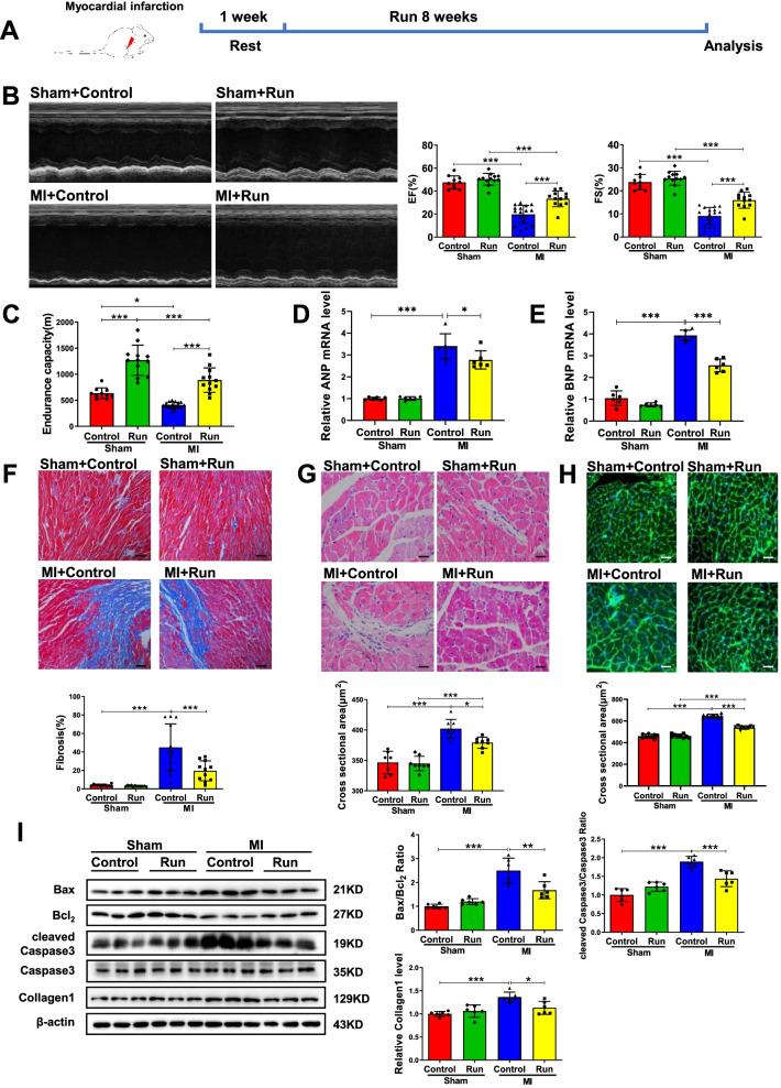
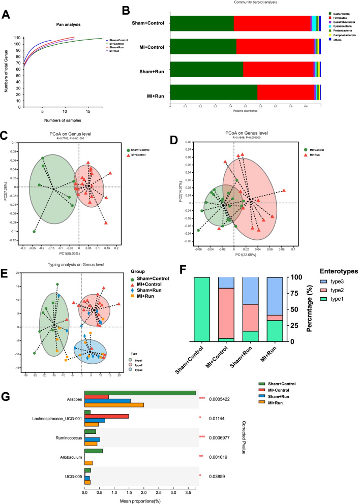
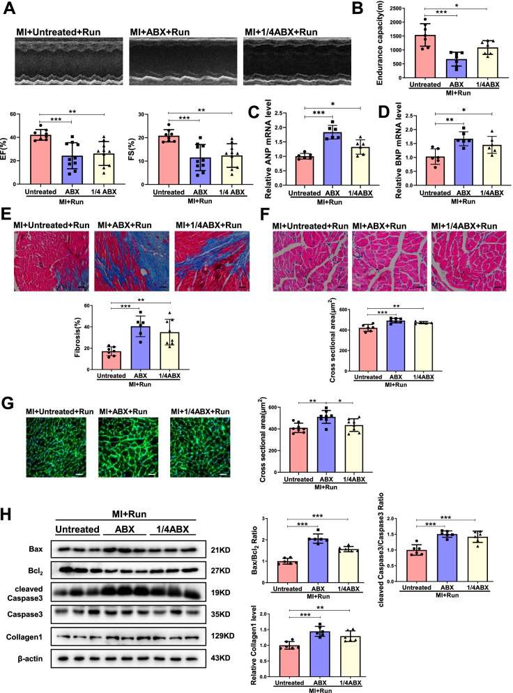
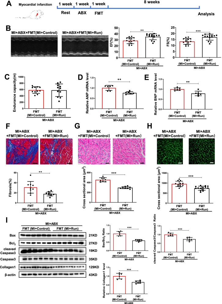
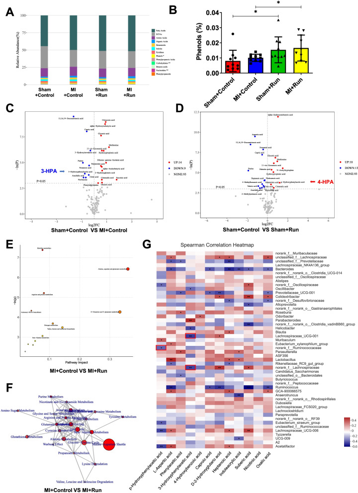
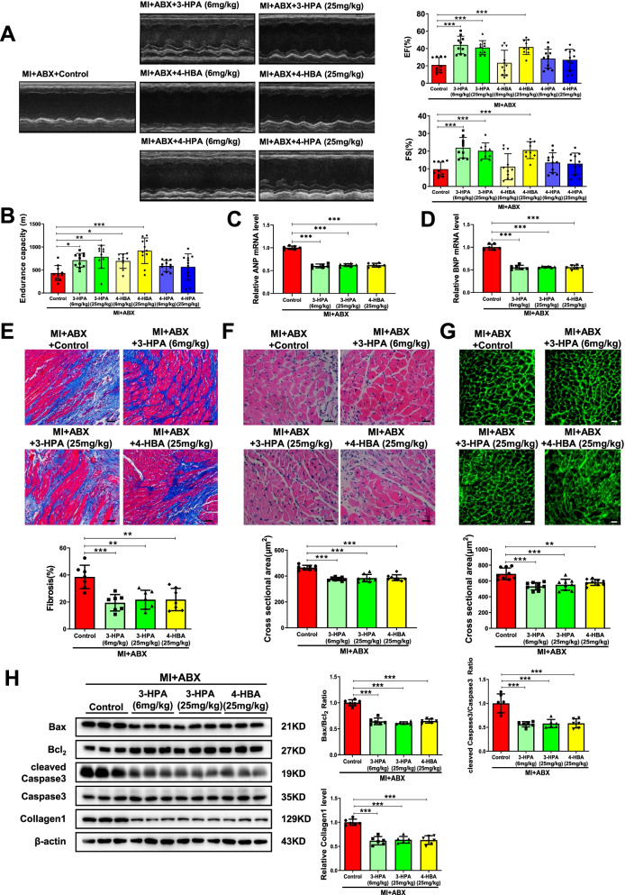
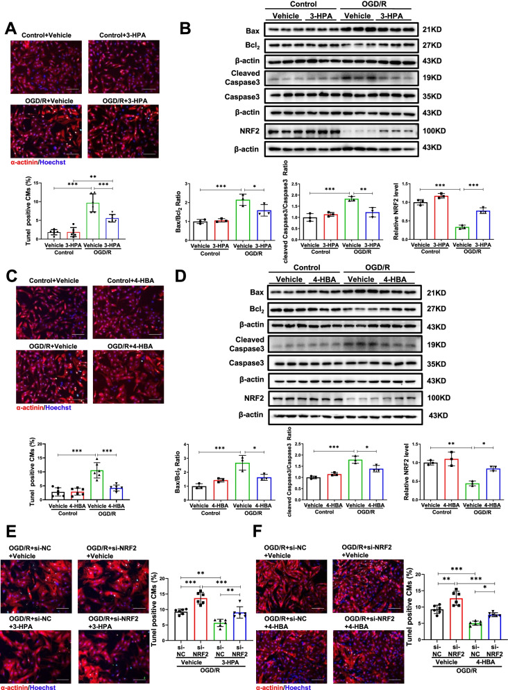
Results: Here, we discovered exercise training ameliorated cardiac dysfunction and changed gut microbial richness and community structure post-MI. Moreover, gut microbiota pre-depletion abolished the protective effects of exercise training in MI mice. Furthermore, mice receiving microbiota transplants from exercised MI mice had better cardiac function compared to mice receiving microbiota transplants from non-exercised MI mice. Mechanistically, we analyzed metabolomics in fecal samples from exercised mice post-MI and identified 3-Hydroxyphenylacetic acid (3-HPA) and 4-Hydroxybenzoic acid (4-HBA), which could be applied individually to protect cardiac dysfunction post-MI and apoptosis through NRF2.

Conclusions: Together, our study provides new insights into the role of gut microbiome in exercise protection to MI, offers opportunities to modulate cardiovascular diseases by exercise, microbiome and gut microbiota-derived 3-HPA and 4-HBA. Video Abstract.

Keywords: Exercise; Gut microbiome; Metabolites; Myocardial infarction; NRF2.


Conflict of interest statement

The authors declare that they have no competing of interests.

Figures

Fig. 1
Fig. 1
Running training protects against cardiac dysfunction after MI. A The schedule of running training after MI. B Running increased ejection fractions (EF) and fractional shortening (FS) post-MI (n = 10:12:17:12). C Running increased endurance capacity post-MI (n = 10:12:17:12). D, E Running decreased cardiac ANP and BNP mRNA expression post-MI (n = 6:6:6:6). F Running decreased cardiac fibrosis post-MI (n = 10:12:12:11). G Running decreased cardiac cross sectional area post-MI by H&E (n = 7:8:8:8). H Running decreased cardiac cross sectional area post-MI by WGA (n = 9:9:9:9). I Running decreased cardiac Bax/Bcl2, cleaved Caspase3/Caspase3 and Collagen1 post-MI (n = 6:6:6:6). Scale bar: 50 μm in F and 25 μm in G and H. Data were represented as mean ± SD. Significant differences were assessed by two-way ANOVA followed by Bonferroni’s multiple comparisons test. *p < 0.05, **p < 0.01, ***p < 0.001
Fig. 2
Fig. 2
Running training changes gut microbial richness and community structure in mice after MI. A Pan analysis for four groups based on genus level (n = 7:18:12:12). B Fecal bacterial community at the phylum level among Sham + control, MI + control, Sham + Run, and MI + Run. C PCoA analysis based on the relative abundance of genus between Sham + control and MI + control groups. D PCoA analysis based on the relative abundance of genus between MI + control and MI + Run groups. E A total of 49 samples were clustered into enterotype 1 (green), enterotype 2 (red), and enterotype 3 (blue) at the genus level. F The percentage of Sham + control, MI + control, Sham + Run, and MI + Run samples distributed in three enterotypes. G Five changed genera among Sham + control, MI + control, Sham + Run, and MI + Run. Significant differences were assessed by ANOSIM analysis in C, D; abund jaccard analysis in E; Wilcoxon rank-sum with FDR in F. *p < 0.05, **p < 0.01, ***p < 0.001
Fig. 3
Fig. 3
Gut microbiota pre-depletion by antibiotics abolishes the protection of running in MI. A Antibiotics (ABX and 1/4 ABX) decreased EF and FS in MI + Run mice (n = 8:10:10). B Antibiotics decreased endurance capacity in MI + Run mice (n = 7:7:7). C, D Antibiotics increased cardiac ANP and BNP mRNA expression in MI + Run mice (n = 6:6:6). E Antibiotics increased cardiac fibrosis in MI + Run mice (n = 7:6:8). F Antibiotics increased cardiac cross sectional area in MI + Run mice by H&E (n = 6:7:5). G Antibiotics increased cardiac cross sectional area in MI + Run mice by WGA (n = 8:8:8). H Antibiotics increased Bax/Bcl2, cleaved Caspase3/Caspase3 and Collagen1 in MI + Run mice (n = 6:6:6). Scale bar: 50 μm in E and 25 μm in F and G. Data were represented as mean ± SD. Significant differences were assessed by one-way ANOVA followed by Bonferroni's multiple comparisons test. *p < 0.05, **p < 0.01, ***p < 0.001
Fig. 4
Fig. 4
FMT recovers the protection of running in MI. A The schedule of FMT after ABX treated. B FMT from MI + Run increased EF and FS compared to FMT from MI + Control post-MI (n = 14:14). C FMT from MI + Run increased endurance capacity compared to FMT from MI + Control post-MI (n = 11:11). D, E FMT from MI + Run decreased cardiac ANP and BNP mRNA expression compared to FMT from MI + control post-MI (n = 6:6). F FMT from MI + Run decreased cardiac fibrosis compared to FMT from MI + control post-MI (n = 9:9). G FMT from MI + Run decreased cardiac cross sectional area compared to FMT from MI + control post-MI by H&E (n = 8:8). H FMT from MI + Run decreased cardiac cross sectional area compared to FMT from MI + control post-MI by WGA (n = 12:14). I FMT from MI + Run decreased Bax/Bcl2, cleaved Caspase3/Caspase3 and collagen1 compared to FMT from MI + Control post-MI (n = 6:6). Scale bar: 50 μm in F and 25 μm in G and H. Data were represented as mean ± SD. Significant differences were assessed by two-tailed student t test. **p < 0.01, ***p < 0.001
Fig. 5
Fig. 5
3-HPA and 4-HPA are identified to be involved in the protection of running in MI. OPLS-DA Model Discrimination based on metabolic profiles in fecal samples. A The relative abundance of each metabolite classes in different groups (n = 10:10:10:10). B The abundance of phenols in different groups. C Volcano plot of univariate statistics across Sham + control group and MI + control group. D Volcano plot of univariate statistics across Sham + control group and Sham + Run group. E Pathway analysis bubble plot by mmu set in MI + Control vs MI + Run. F Network for statistically significant changed pathways in MI + control vs. MI + Run. G The relationship between metabolites and the top 50 genera in four groups was estimated by Spearman’s correlation analysis. Those with low correlated (|r|< 0.1) were not shown. Genera and cardiac function index were distinguished as positive (red) and negative (blue) correlation. *p < 0.05; **p < 0.01; ***p < 0.001
Fig. 6
Fig. 6
3-HPA and 4-HBA protect against cardiac dysfunction after MI. A 3-HPA and 4-HBA increased EF and FS post-MI (n = 10:10:9:11:9:11:11). B 3-HPA and 4-HBA increased endurance capacity post-MI (n = 10:11:10:10:11:11:11). C, D 3-HPA and 4-HBA decreased cardiac ANP and BNP mRNA expression post-MI (n = 6:6:6:6). E 3-HPA and 4-HBA decreased cardiac fibrosis post-MI (n = 7:7:7:7). F 3-HPA and 4-HBA decreased cardiac cross sectional area post-MI by H&E (n = 8:8:8:8). G 3-HPA and 4-HBA decreased cardiac cross sectional area post-MI by WGA (n = 9:9:9:9). H 3-HPA and 4-HBA decreased Bax/Bcl2, cleaved Caspase3/Caspase3 and Collagen1 post-MI (n = 6:6:6:6). Scale bar: 50 μm in E and 25 μm in F and G. Data were represented as mean ± SD. Significant differences were assessed by one-way ANOVA followed by Bonferroni's multiple comparisons test. *p < 0.05, **p < 0.01, ***p < 0.001
Fig. 7
Fig. 7
3-HPA and 4-HBA mediate cardiac protection through activating NRF2. A, B 3-HPA (100 μM, 24 h) reduced the percentage of TUNEL staining positive cardiomyocytes (n = 6:6:6:6), Bax/Bcl2, cleaved Caspase3/Caspase3, NRF2 (n = 3:3:3:3) in NRCMs under oxygen glucose deprivation/reperfusion (OGD/R). C, D 4-HBA (100 μM, 24 h) decreased the percentage of TUNEL staining positive cardiomyocytes (n = 6:6:6:6), Bax/Bcl2, cleaved Caspase3/Caspase3, NRF2 (n = 3:3:3:3) in NRCMs under OGD/R. E, F Inhibition of NRF2 increased the percentage of TUNEL staining positive cardiomyocytes decreased by 3-HPA and 4-HBA in NRCMs under OGD/R (n = 6:6:6:6). Scale bar: 100 μm. Data were represented as mean ± SD. Significant differences were assessed by two-way ANOVA followed by Bonferroni's multiple comparisons test. *p < 0.05, **p < 0.01, ***p < 0.001 versus respective control

References

    1. Degnan PH, Barry NA, Mok KC, Taga ME, Goodman AL. Human gut microbes use multiple transporters to distinguish vitamin B(1)(2) analogs and compete in the gut. Cell Host Microbe. 2014;15(1):47–57. doi: 10.1016/j.chom.2013.12.007. - DOI - PMC - PubMed
    1. Nicholson JK, Holmes E, Wilson ID. Gut microorganisms, mammalian metabolism and personalized health care. Nat Rev Microbiol. 2005;3(5):431–438. doi: 10.1038/nrmicro1152. - DOI - PubMed
    1. Liu R, Hong J, Xu X, Feng Q, Zhang D, Gu Y, Shi J, Zhao S, Liu W, Wang X, et al. Gut microbiome and serum metabolome alterations in obesity and after weight-loss intervention. Nat Med. 2017;23(7):859–868. doi: 10.1038/nm.4358. - DOI - PubMed
    1. Cani PD. Human gut microbiome: hopes, threats and promises. Gut. 2018;67(9):1716–1725. doi: 10.1136/gutjnl-2018-316723. - DOI - PMC - PubMed
    1. Loomba R, Seguritan V, Li W, Long T, Klitgord N, Bhatt A, Dulai PS, Caussy C, Bettencourt R, Highlander SK, et al. Gut microbiome-based metagenomic signature for non-invasive detection of advanced fibrosis in human nonalcoholic fatty liver disease. Cell Metab. 2017;25(5):1054–1062 e1055. doi: 10.1016/j.cmet.2017.04.001. - DOI - PMC - PubMed

Publication types