

White-centered retinal hemorrhage revealing acute leukemia: A case report
- PMID: 35638035
- PMCID: PMC9142656
- DOI: 10.1016/j.amsu.2022.103632
White-centered retinal hemorrhage revealing acute leukemia: A case report
Abstract
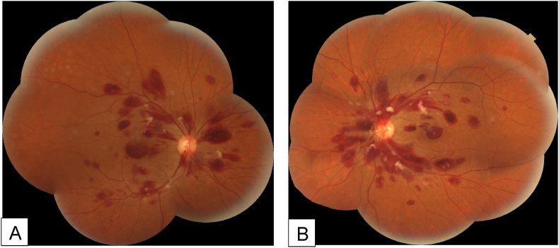
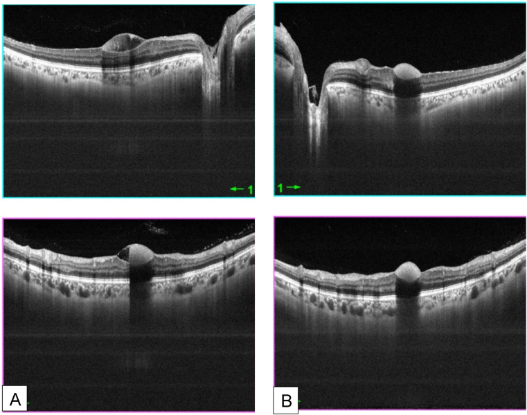
Retinal manifestations have been described as an inaugural manifestation of acute leukemia. Retinal hemorrhage, and in particular white-centered hemorrhages are among the most frequently observed signs. We report here the case of a 34-year-old Caucasian man with no medical history who presented to our emergency department with a decrease in visual acuity associated with asthenia. Ophthalmological examination revealed bilateral white-centered hemorrhages. The etiological assessment confirmed the diagnosis of acute myeloid leukemia. Whenever Roth spots are found in fundus examination, a complete ophthalmological examination along with a wide etiological investigation must be conducted.
Keywords: Acute myeloid leukemia; Case report; Roth spots; White-centered hemorrhage.
© 2022 The Authors.
Conflict of interest statement
The following authors have no financial disclosures: (Nadia Ben Abdessalem, Nesrine Zaafrane, Atf Ben Abderrazek, Ahmed Jabri, Anis Mahjoub, Chiraz Ben Youssef, Hachemi Mahjoub, Fathi Krifa, Ahmed Mahjoub).
Figures
Similar articles
-
White-centred retinal hemorrhage revealing acute leukemia.Tunis Med. 2019 Jun;97(6):822-825. Tunis Med. 2019. PMID: 31872415
-
Highlights of ophthalmological manifestations in newly diagnosed acute leukemia: a correlation with hematological parameters.Ann Hematol. 2024 Sep;103(9):3519-3533. doi: 10.1007/s00277-024-05861-2. Epub 2024 Jul 10. Ann Hematol. 2024. PMID: 38985179 Free PMC article.
-
Visual Disturbance as the first Symptom of Chronic Myeloid Leukemia.Middle East Afr J Ophthalmol. 2011 Oct;18(4):336-8. doi: 10.4103/0974-9233.90143. Middle East Afr J Ophthalmol. 2011. PMID: 22224030 Free PMC article.
-
Roth spots seen on ophthalmoscopy: diseases with which they may be associated.Conn Med. 1995 May;59(5):271-3. Conn Med. 1995. PMID: 7600798 Review.
-
Bilateral retinal hemorrhages from megaloblastic anemia: case report and review of literature.Ann Ophthalmol. 1992 Mar;24(3):86-90. Ann Ophthalmol. 1992. PMID: 1570927 Review.
Cited by
-
Ophthalmic Manifestations of Newly Diagnosed Acute Leukemia Patients in a Tunisian Cohort.Clin Ophthalmol. 2022 Oct 14;16:3425-3435. doi: 10.2147/OPTH.S365648. eCollection 2022. Clin Ophthalmol. 2022. PMID: 36249442 Free PMC article.
References
-
- Chaabani L., Doulami K. White-centred retinal hemorrhage revealing acute leukemia. Tunis. Med. 2019 Jun;97(6):822–825. PMID: 31872415. - PubMed
-
- abu el-Asrar A.M., al-Momen A.K., Kangave D., Harakati M.S., Ajarim D.S. Correlation of fundus lesions and hematologic findings in leukemic retinopathy. Eur. J. Ophthalmol. 1996 Apr-Jun;6(2):167–172. PMID: 8823591. - PubMed
-
- Agha R.A., Franchi T., Sohrabi C., Mathew G., for the SCARE Group The SCARE 2020 guideline: updating consensus Surgical CAse REport (SCARE) guidelines. Int. J. Surg. 2020;84:226–230. - PubMed
Publication types
LinkOut - more resources
Full Text Sources
