. 2022 Jun 28;13(3):e0130022.
doi: 10.1128/mbio.01300-22. Epub 2022 May 31.

The Deubiquitinase USP29 Promotes SARS-CoV-2 Virulence by Preventing Proteasome Degradation of ORF9b

Affiliations

The Deubiquitinase USP29 Promotes SARS-CoV-2 Virulence by Preventing Proteasome Degradation of ORF9b

Wenying Gao et al. mBio. .

Abstract

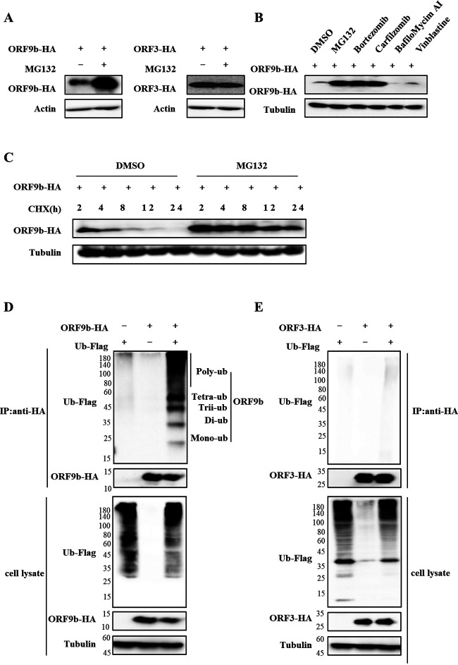
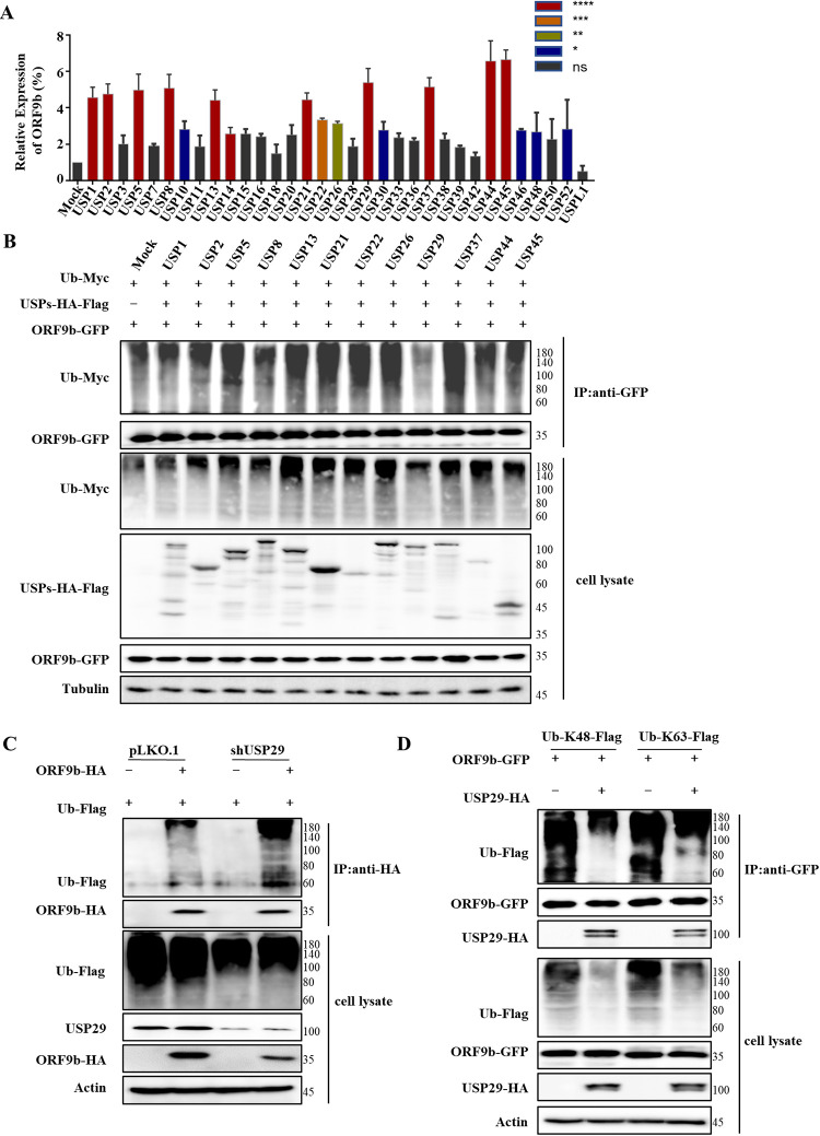
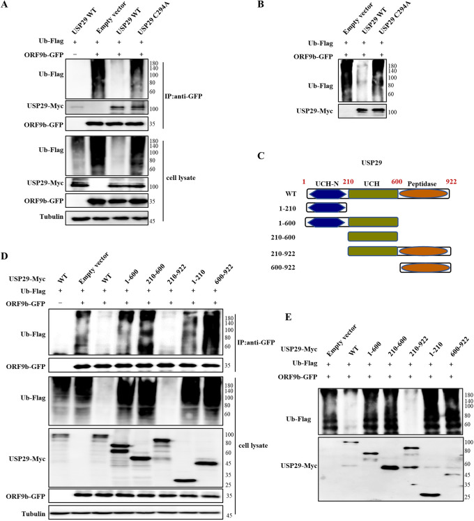
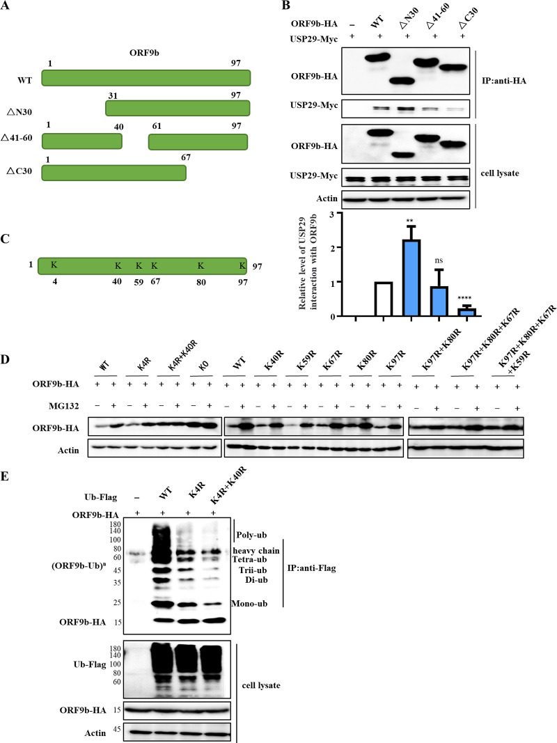
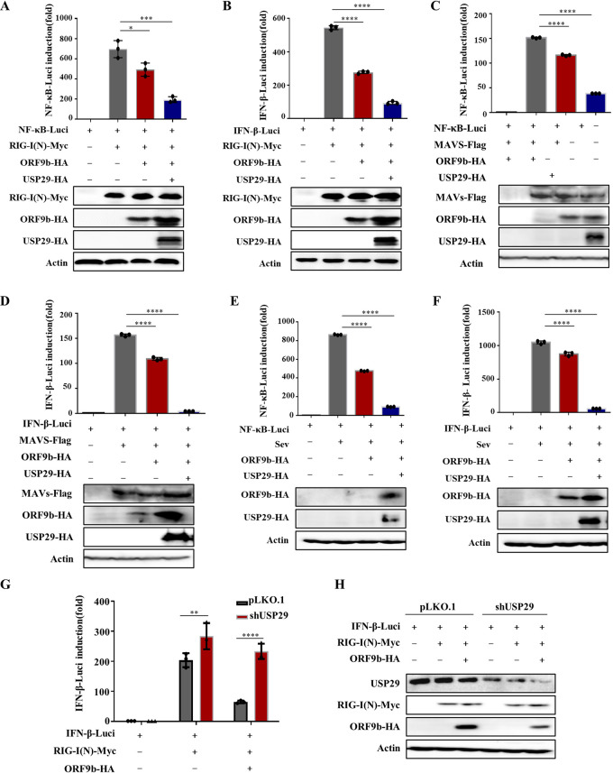
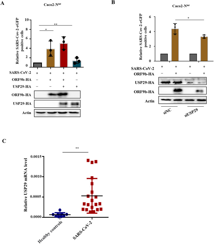
Ubiquitin signaling is essential for immunity to restrict pathogen proliferation. Due to its enormous impact on human health and the global economy, intensive efforts have been invested in studying severe acute respiratory syndrome coronavirus 2 (SARS-CoV-2) and its interactions with hosts. However, the role of the ubiquitin network in pathogenicity has not yet been explored. Here, we found that ORF9b of SARS-CoV-2 is ubiquitinated on Lys-4 and Lys-40 by unknown E3 ubiquitin ligases and is degraded by the ubiquitin proteasomal system. Importantly, we identified USP29 as a host factor that prevents ORF9b ubiquitination and subsequent degradation. USP29 interacts with the carboxyl end of ORF9b and removes ubiquitin chains from the protein, thereby inhibiting type I interferon (IFN) induction and NF-κB activation. We also found that ORF9b stabilization by USP29 enhanced the virulence of VSV-eGFP and transcription and replication-competent SARS-CoV-2 virus-like-particles (trVLP). Moreover, we observed that the mRNA level of USP29 in SARS-CoV-2 patients was higher than that in healthy people. Our findings provide important evidence indicating that targeting USP29 may effectively combat SARS-CoV-2 infection. IMPORTANCE Coronavirus disease 2019 (COVID-19) is a current global health threat caused by SARS-CoV-2. The innate immune response such as type I IFN (IFN-I) is the first line of host defense against viral infections, whereas SARS-CoV-2 proteins antagonize IFN-I production through distinct mechanisms. Among them, ORF9b inhibits the canonical IκB kinase alpha (IKKɑ)/β/γ-NF-κB signaling and subsequent IFN production; therefore, discovering the regulation of ORF9b by the host might help develop a novel antiviral strategy. Posttranslational modification of proteins by ubiquitination regulates many biological processes, including viral infections. Here, we report that ORF9b is ubiquitinated and degraded through the proteasome pathway, whereas deubiquitinase USP29 deubiquitinates ORF9b and prevents its degradation, resulting in the enhancement of ORF9b-mediated inhibition of IFN-I and NF-κB activation and the enhancement of virulence of VSV-eGFP and SARS-CoV-2 trVLP.

Keywords: ORF9b; SARS-CoV-2; USP29; degradation; deubiquitination.


Conflict of interest statement

The authors declare no conflict of interest.

Figures

FIG 1
FIG 1
ORF9b is ubiquitinated and degraded through the proteasomal pathway. (A) Proteasomal inhibitor MG132 increased the expression of ORF9b but not ORF3a. (B) Proteasomal inhibitors, but not other inhibitors, increased ORF9b expression. The ORF9b-HA-tag expression vector was transfected into HEK293T cells, and then the cells were treated with 10 μM MG132, Bortezomib, Carfilzomib, BafiloMycim AI, Vinblastine, or DMSO for 12 h prior to harvest. The cell lysates were analyzed using immunoblot (IB). (C) MG132 stabilized ORF9b expression in cells treated with CHX. ORF9b-HA was transfected into HEK293T cells for 24 h, and the cells were treated with or without 10 μM MG132 for 10 h, then 50 μg/mL cycloheximide (CHX) was added. The cells were harvested at different time points, and their lysates were analyzed. ORF9b (D) but not ORF3 (E) are ubiquitinated. HEK293T cells transfected with ORF9b-HA or ORF3-HA plus Ub-Flag or the empty vector were treated with 10 mM MG132 for 12 h prior to harvest. Co-IP (with anti-HA) and IB analysis were performed.
FIG 2
FIG 2
USP29 prevents ORF9b degradation by ubiquitination. (A) Effect of 33 deubiquitinase USPs on ORF9b expression. ORF9b-HA and USPs-HA-Flag or control vector were cotransfected into HEK293T cells. Cells were harvested at 48 h posttransfection and analyzed using IB. Quantification of ORF9b expression was performed using ImageJ2X, its expression in cells cotransfected with ORF9b, and the control vector was set to 100%. Statistical significance was analyzed using two-sided unpaired t tests (NS, not significant; *, P < 0.05; **, P < 0.01; ***, P < 0.001; ****, P < 0.0001). (B) USP29 deubiquitinated ORF9b. HEK293T cells were transfected with ORF9b-GFP, Ub-Myc, and USPs-HA-Flag, and treated with 10 mM MG132 for 12 h prior to harvest. Cell lysates were immunoprecipitated with anti-GFP antibodies conjugated to agarose beads. Cell lysates and precipitated samples were analyzed using IB with the corresponding antibodies. (C) USP29 silencing reduced deubiquitination of ORF9b. ORF9b ubiquitination in USP29 silencing and control cells were analyzed using Co-IP (with anti-Flag) and IB. (D) USP29 could cleave both K48- and K63-linked ubiquitin chains of ORF9b.
FIG 3
FIG 3
The enzymatic activity and the C-terminus of USP29 are required for ORF9b deubiquitination. (A) USP29 deubiquitinase-deficient C294A mutant could not deubiquitinate ORF9b in vivo. HEK293T cells transfected with the indicated expression vectors were treated with 10 mM MG132 for 12 h prior to harvest. Cell lysates were immunoprecipitated by protein G agarose beads conjugated with anti-GFP antibodies. Cell lysates and precipitated samples were analyzed using IB with the corresponding antibodies. (B) In vitro deubiquitination assay. Ubiquitinated ORF9b was immunoprecipitated from HEK293T cells transfected with ORF9b-GFP and Ub-Flag by anti-GFP antibody-conjugated protein G agarose beads. USP29 or its mutant was immunoprecipitated from HEK293T cells overexpressing USP29-Myc or its mutant using anti-Myc antibody-conjugated protein G agarose. Ubiquitinated ORF9b was incubated with purified USP29 or its mutant in deubiquitination buffer for 12 h at 37°C, and then was analyzed using IB. (C) The schematic represents USP29 WT and mutants. The C-terminus of USP29 is required for ORF9b deubiquitination in vivo (D) and in vitro (E) assays. The experimental process was similar to A and B.
FIG 4
FIG 4
The ubiquitinated sites of ORF9b. (A) The schematic represents ORF9b WT and mutants used in the study. (B) The interaction between ORF9b and its mutants with USP29. HEK293T cells cotransfected with USP29-Myc and ORF9b-HA WT or mutants were subjected to Co-IP with anti-HA agarose beads. The immunoprecipitates and lysates were analyzed using IB. Quantification of USP29 interaction with ORF9b was performed using ImageJ2X. Data were normalized to pulldown ORF9b WT or mutations, respectively. The interaction of ORF9b WT and USP29 was set 1. Statistical significance was analyzed using two-sided unpaired t tests (NS, not significant; **, P < 0.01; ****, P < 0.0001). (C) Schema showing the lysines (K) of SARS-CoV-2 ORF9b. (D) The stability of ORF9b lysine mutants. (E) The ubiquitination of ORF9b lysine mutants. HEK293T cells transfected with ORF9b WT or mutants plus ubiquitin-Flag were treated with 10 mM MG132 for 12 h prior to harvest. Cell lysates were immunoprecipitated by protein G agarose beads conjugated with anti-Flag antibodies. Cell lysates and precipitated samples were analyzed using IB.
FIG 5
FIG 5
USP29 enhances ORF9b-mediated IFN and NF-κB inhibition by increasing ORF9b expression. (A–D) USP29 promotes ORF9b inhibition on RIG-N or MAVS to activate IFN-β/NF-κB promoters. HEK293T cells were transfected with an IFN-β/NF-κB promoter Luciferase reporter plasmid, renilla Luciferase reporter plasmid, or ORF9b-HA, along with a control plasmid or USP29-HA for 24 h. Then Luciferase reporter activities were induced by transfection of RIG-I(N)-Myc or MAVS-Flag expressing vectors into HEK293T cells for another 24 h. Cell lysate samples were analyzed using IB with the corresponding antibodies (lower panels). NF-κB-Luc (A, C) and IFN-β-Luc (B, D) reporter activities are normalized to renilla Luciferase and shown as fold induction. (E–F) USP29 promotes ORF9b inhibition of activation of IFN-β/NF-κB promoters by SeV infection. HEK293T cells were transfected with an IFN-β/NF-κB reporter plasmid, renilla reporter plasmid, or ORF9b-HA, along with a control plasmid or USP29-HA for 24 h. Then, Luciferase reporter activities were induced by SeV infection for 12 h. Cell lysate samples were analyzed using IB with the corresponding antibodies (lower panels). NF-κB-Luc (E) and IFN-β-Luc (F) reporter activities are normalized to that of renilla Luciferase and shown as fold induction. (G–H) Knockdown of USP29 reduced ORF9b-mediated IFN inhibition. USP29 silencing or control HEK293T cells were transfected with IFN-β reporter plasmid, renilla reporter plasmid, ORF9b-HA, control plasmid, or USP29-HA for 24 h. Then Luciferase reporter activities were induced by transfection of RIG-I(N)-Myc into USP29 silenced or control HEK293T cells for 24 h. (G) IFN-β-Luc reporter activities are normalized to renilla Luciferase and shown as fold induction. (H) Cell lysate samples were analyzed using IB with the corresponding antibodies. Data are represented as means ± SDs calculated from three independent experiments (****, P < 0.0001).
FIG 6
FIG 6
USP29 increases VSV viral infectivity through stabilizing ORF9b. (A) Schematic presentation of assessment of USP29 increases VSV viral infectivity through stabilizing ORF9b. (B–E) At 24 h posttransfection of empty vector or ORF9b and USP29-HA expressing plasmids, HEK293T cells were infected with VSV-eGFP for 12 h. (B) Fluorescent images were taken to examine VSV-eGFP proliferation. (C) eGFP positive cells were analyzed using flow cytometry, and the mRNA expression of IFN-β was determined by RT-qPCR (D). (E) Cell lysate samples were analyzed using IB with the corresponding antibodies.
FIG 7
FIG 7
USP29 increases SARS-CoV-2 trVLP infectivity through stabilizing ORF9b. (A) Caco-2-Nint cells were transfected with ORF9b-HA plus control plasmid or USP29-HA for 24 h. Then cells were infected with SARS-CoV-2 trVLPs at a multiplicity of infection (MOI) of 0.1 for 48 h. The infection was analyzed using flow cytometry to detect eGFP-positive cells (top). Protein expression was analyzed using IB (bottom). (B) USP29 silencing reduced SARS-CoV-2 trVLPs infectivity by reducing ORF9b expression. Caco-2-Nint cells were transfected with ORF9b-HA for 24 h. Infection and IB were conducted as described in A. (C) Total mRNA was extracted from PBMC cells of a healthy person or recovered SARS-CoV-2 patients. USP29 mRNA levels were determined using RT-qPCR. Statistical significance was analyzed using two-sided unpaired t tests (NS, not significant; *, P < 0.05; **, P < 0.01; ***, P < 0.001; ****, P < 0.0001).

Similar articles

Cited by

References

    1. Redondo N, Zaldivar-Lopez S, Garrido JJ, Montoya M. 2021. SARS-CoV-2 accessory proteins in viral pathogenesis: knowns and unknowns. Front Immunol 12:708264. doi:10.3389/fimmu.2021.708264. - DOI - PMC - PubMed
    1. Han L, Zhuang MW, Deng J, Zheng Y, Zhang J, Nan ML, Zhang XJ, Gao C, Wang PH. 2021. SARS-CoV-2 ORF9b antagonizes type I and III interferons by targeting multiple components of the RIG-I/MDA-5-MAVS, TLR3-TRIF, and cGAS-STING signaling pathways. J Med Virol 93:5376–5389. doi:10.1002/jmv.27050. - DOI - PMC - PubMed
    1. Wu J, Shi Y, Pan X, Wu S, Hou R, Zhang Y, Zhong T, Tang H, Du W, Wang L, Wo J, Mu J, Qiu Y, Yang K, Zhang LK, Ye BC, Qi N. 2021. SARS-CoV-2 ORF9b inhibits RIG-I-MAVS antiviral signaling by interrupting K63-linked ubiquitination of NEMO. Cell Rep 34:108761. doi:10.1016/j.celrep.2021.108761. - DOI - PMC - PubMed
    1. Gao X, Zhu K, Qin B, Olieric V, Wang M, Cui S. 2021. Crystal structure of SARS-CoV-2 Orf9b in complex with human TOM70 suggests unusual virus-host interactions. Nat Commun 12:2843. doi:10.1038/s41467-021-23118-8. - DOI - PMC - PubMed
    1. Hrecka K, Hao C, Gierszewska M, Swanson SK, Kesik-Brodacka M, Srivastava S, Florens L, Washburn MP, Skowronski J. 2011. Vpx relieves inhibition of HIV-1 infection of macrophages mediated by the SAMHD1 protein. Nature 474:658–661. doi:10.1038/nature10195. - DOI - PMC - PubMed

Publication types